 Movie
Movie Controller

[English] 日本語
 Yorodumi
- PDB-5j3d: Crystal structure of human Fab 14N4 in complex with post-fusion RSV F -
+ Open data

- Basic information
| Entry | Database: PDB / ID: 5j3d | ||||||
|---|---|---|---|---|---|---|---|
| Title | Crystal structure of human Fab 14N4 in complex with post-fusion RSV F | ||||||
 Components Components |
| ||||||
 Keywords Keywords | IMMUNE SYSTEM / antibody / virus / complex | ||||||
| Function / homology |  Function and homology information Function and homology informationsymbiont-mediated induction of syncytium formation / Translation of respiratory syncytial virus mRNAs / RSV-host interactions / Assembly and release of respiratory syncytial virus (RSV) virions / Maturation of hRSV A proteins / Respiratory syncytial virus (RSV) attachment and entry / host cell Golgi membrane / entry receptor-mediated virion attachment to host cell / fusion of virus membrane with host plasma membrane / viral envelope ...symbiont-mediated induction of syncytium formation / Translation of respiratory syncytial virus mRNAs / RSV-host interactions / Assembly and release of respiratory syncytial virus (RSV) virions / Maturation of hRSV A proteins / Respiratory syncytial virus (RSV) attachment and entry / host cell Golgi membrane / entry receptor-mediated virion attachment to host cell / fusion of virus membrane with host plasma membrane / viral envelope / symbiont entry into host cell / host cell plasma membrane / virion membrane / identical protein binding / plasma membrane Similarity search - Function | ||||||
| Biological species |  Homo sapiens (human) Homo sapiens (human) Human respiratory syncytial virus A Human respiratory syncytial virus A | ||||||
| Method |  X-RAY DIFFRACTION / SYNCHROTRON / MOLECULAR REPLACEMENT / Resolution: 4.077 Å X-RAY DIFFRACTION / SYNCHROTRON / MOLECULAR REPLACEMENT / Resolution: 4.077 Å | ||||||
 Authors Authors | Mousa, J.J. / Crowe, J.E. | ||||||
 Citation Citation | Journal: Proc.Natl.Acad.Sci.USA / Year: 2016 Title: Structural basis for nonneutralizing antibody competition at antigenic site II of the respiratory syncytial virus fusion protein. Authors: Mousa, J.J. / Sauer, M.F. / Sevy, A.M. / Finn, J.A. / Bates, J.T. / Alvarado, G. / King, H.G. / Loerinc, L.B. / Fong, R.H. / Doranz, B.J. / Correia, B.E. / Kalyuzhniy, O. / Wen, X. / ...Authors: Mousa, J.J. / Sauer, M.F. / Sevy, A.M. / Finn, J.A. / Bates, J.T. / Alvarado, G. / King, H.G. / Loerinc, L.B. / Fong, R.H. / Doranz, B.J. / Correia, B.E. / Kalyuzhniy, O. / Wen, X. / Jardetzky, T.S. / Schief, W.R. / Ohi, M.D. / Meiler, J. / Crowe, J.E. | ||||||
| History |
|
- Structure visualization
| Structure viewer | Molecule: MolmilJmol/JSmol |
|---|
- Downloads & links
-Download
| PDBx/mmCIF format | 5j3d.cif.gz | 501.9 KB | Display | PDBx/mmCIF format |
|---|---|---|---|---|
| PDB format | pdb5j3d.ent.gz | 409.4 KB | Display | PDB format |
| PDBx/mmJSON format | 5j3d.json.gz | Tree view | PDBx/mmJSON format | |
| Others |  Other downloads Other downloads |
-Validation report
| Arichive directory | https://data.pdbj.org/pub/pdb/validation_reports/j3/5j3dftp://data.pdbj.org/pub/pdb/validation_reports/j3/5j3d | HTTPS FTP |
|---|
-Related structure data
| Related structure data |  5itbC 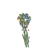 3rrrS C: citing same article ( S: Starting model for refinement |
|---|---|
| Similar structure data |


-Links
 PDBj
PDBj


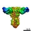
- Assembly
| Deposited unit | 
| ||||||||
|---|---|---|---|---|---|---|---|---|---|
| 1 |
| ||||||||
| Unit cell |
|
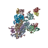
-Components
| #1: Antibody | Mass: 23597.490 Da / Num. of mol.: 3 Source method: isolated from a genetically manipulated source Source: (gene. exp.) Homo sapiens (human) / Production host: Homo sapiens (human)#2: Antibody | Mass: 24001.520 Da / Num. of mol.: 3 Source method: isolated from a genetically manipulated source Source: (gene. exp.) Homo sapiens (human) / Production host: Homo sapiens (human)#3: Protein | Mass: 8301.455 Da / Num. of mol.: 3 / Fragment: UNP residues 26-98 Source method: isolated from a genetically manipulated source Source: (gene. exp.) Human respiratory syncytial virus A / Production host: Homo sapiens (human) / References: UniProt: P03420#4: Protein | Mass: 43782.762 Da / Num. of mol.: 3 / Fragment: UNP residues 147-513 Source method: isolated from a genetically manipulated source Source: (gene. exp.) Human respiratory syncytial virus A / Production host: Homo sapiens (human) / References: UniProt: P03420Has protein modification | Y | |
|---|
-Experimental details
-Experiment
| Experiment | Method: X-RAY DIFFRACTION / Number of used crystals: 1 |
|---|
- Sample preparation
| Crystal | Density Matthews: 5.17 Å3/Da / Density % sol: 76.23 % |
|---|---|
| Crystal grow | Temperature: 293 K / Method: vapor diffusion / Details: 2 M ammonium sulfate, 5% 2-propanol |
-Data collection
| Diffraction | Mean temperature: 80 K |
|---|---|
| Diffraction source | Source: SYNCHROTRON / Site: APS  / Beamline: 21-ID-F / Wavelength: 0.97872 Å / Beamline: 21-ID-F / Wavelength: 0.97872 Å |
| Detector | Type: RAYONIX MX-225 / Detector: CCD / Date: Dec 11, 2015 |
| Radiation | Protocol: SINGLE WAVELENGTH / Monochromatic (M) / Laue (L): M / Scattering type: x-ray |
| Radiation wavelength | Wavelength: 0.97872 Å / Relative weight: 1 |
| Reflection | Resolution: 4.077→49.5 Å / Num. obs: 91490 / % possible obs: 98.1 % / Redundancy: 8.3 % / CC1/2: 0.987 / Rmerge(I) obs: 0.296 / Net I/σ(I): 5.5 |
| Reflection shell | Resolution: 4.077→4.25 Å / Redundancy: 8.5 % / Rmerge(I) obs: 1.191 / Mean I/σ(I) obs: 1.7 / CC1/2: 0.511 / % possible all: 98.7 |
- Processing
| Software |
| ||||||||||||||||||||||||||||||||||||||||||||||||||||||||||||||||||||||||||||||||||||||||||||||||||||||||||||||||||||||||||||||||||||||||||||||||||||||||||||||||||||||||||||||||||||||||||||||||||||
|---|---|---|---|---|---|---|---|---|---|---|---|---|---|---|---|---|---|---|---|---|---|---|---|---|---|---|---|---|---|---|---|---|---|---|---|---|---|---|---|---|---|---|---|---|---|---|---|---|---|---|---|---|---|---|---|---|---|---|---|---|---|---|---|---|---|---|---|---|---|---|---|---|---|---|---|---|---|---|---|---|---|---|---|---|---|---|---|---|---|---|---|---|---|---|---|---|---|---|---|---|---|---|---|---|---|---|---|---|---|---|---|---|---|---|---|---|---|---|---|---|---|---|---|---|---|---|---|---|---|---|---|---|---|---|---|---|---|---|---|---|---|---|---|---|---|---|---|---|---|---|---|---|---|---|---|---|---|---|---|---|---|---|---|---|---|---|---|---|---|---|---|---|---|---|---|---|---|---|---|---|---|---|---|---|---|---|---|---|---|---|---|---|---|---|---|---|---|
| Refinement | Method to determine structure: MOLECULAR REPLACEMENT Starting model: 3RRR Resolution: 4.077→49.5 Å / SU ML: 0.54 / Cross valid method: FREE R-VALUE / σ(F): 1.33 / Phase error: 26.91 / Stereochemistry target values: ML
| ||||||||||||||||||||||||||||||||||||||||||||||||||||||||||||||||||||||||||||||||||||||||||||||||||||||||||||||||||||||||||||||||||||||||||||||||||||||||||||||||||||||||||||||||||||||||||||||||||||
| Solvent computation | Shrinkage radii: 0.9 Å / VDW probe radii: 1.11 Å / Solvent model: FLAT BULK SOLVENT MODEL | ||||||||||||||||||||||||||||||||||||||||||||||||||||||||||||||||||||||||||||||||||||||||||||||||||||||||||||||||||||||||||||||||||||||||||||||||||||||||||||||||||||||||||||||||||||||||||||||||||||
| Refinement step | Cycle: LAST / Resolution: 4.077→49.5 Å
| ||||||||||||||||||||||||||||||||||||||||||||||||||||||||||||||||||||||||||||||||||||||||||||||||||||||||||||||||||||||||||||||||||||||||||||||||||||||||||||||||||||||||||||||||||||||||||||||||||||
| Refine LS restraints |
| ||||||||||||||||||||||||||||||||||||||||||||||||||||||||||||||||||||||||||||||||||||||||||||||||||||||||||||||||||||||||||||||||||||||||||||||||||||||||||||||||||||||||||||||||||||||||||||||||||||
| LS refinement shell |
|
