 Movie
Movie Controller

[English] 日本語
 Yorodumi
- PDB-4ydx: Crystal structure of cisplatin bound to a human copper chaperone ... -
+ Open data

- Basic information
| Entry | Database: PDB / ID: 4ydx | ||||||
|---|---|---|---|---|---|---|---|
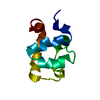
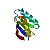
| Title | Crystal structure of cisplatin bound to a human copper chaperone (monomer) - new refinement | ||||||
 Components Components | Copper transport protein ATOX1 | ||||||
 Keywords Keywords | METAL TRANSPORT / Re-refinement of 3iwl / cisplatin / platinum / Metal-binding | ||||||
| Function / homology |  Function and homology information Function and homology informationmetallochaperone activity / Ion influx/efflux at host-pathogen interface / copper-dependent protein binding / copper chaperone activity / copper ion export / copper ion transport / Detoxification of Reactive Oxygen Species / cuprous ion binding / intracellular copper ion homeostasis / ATPase binding ...metallochaperone activity / Ion influx/efflux at host-pathogen interface / copper-dependent protein binding / copper chaperone activity / copper ion export / copper ion transport / Detoxification of Reactive Oxygen Species / cuprous ion binding / intracellular copper ion homeostasis / ATPase binding / response to oxidative stress / copper ion binding / negative regulation of apoptotic process / cytosol Similarity search - Function | ||||||
| Biological species |  Homo sapiens (human) Homo sapiens (human) | ||||||
| Method |  X-RAY DIFFRACTION / SYNCHROTRON / FOURIER SYNTHESIS / Resolution: 1.602 Å X-RAY DIFFRACTION / SYNCHROTRON / FOURIER SYNTHESIS / Resolution: 1.602 Å | ||||||
 Authors Authors | Shabalin, I.G. / Boal, A.K. / Dauter, Z. / Jaskolski, M. / Minor, W. / Rosenzweig, A.C. / Wlodawer, A. | ||||||
| Funding support |  United States, 1items United States, 1items
| ||||||
 Citation Citation | Journal: Acta Crystallogr.,Sect.D / Year: 2015 Title: Crystallography and chemistry should always go together: a cautionary tale of protein complexes with cisplatin and carboplatin. Authors: Shabalin, I. / Dauter, Z. / Jaskolski, M. / Minor, W. / Wlodawer, A. #1: Journal: J.Am.Chem.Soc. / Year: 2009Title: Crystal structures of cisplatin bound to a human copper chaperone. Authors: Boal, A.K. / Rosenzweig, A.C. | ||||||
| History |
|
- Structure visualization
| Structure viewer | Molecule: MolmilJmol/JSmol |
|---|
- Downloads & links
-Download
| PDBx/mmCIF format | 4ydx.cif.gz | 45.7 KB | Display | PDBx/mmCIF format |
|---|---|---|---|---|
| PDB format | pdb4ydx.ent.gz | 30.9 KB | Display | PDB format |
| PDBx/mmJSON format | 4ydx.json.gz | Tree view | PDBx/mmJSON format | |
| Others |  Other downloads Other downloads |
-Validation report
| Arichive directory | https://data.pdbj.org/pub/pdb/validation_reports/yd/4ydxftp://data.pdbj.org/pub/pdb/validation_reports/yd/4ydx | HTTPS FTP |
|---|
-Related structure data
| Related structure data |  4yeaC  4yemC 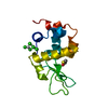 4yenC 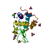 4yeoC  3iwlS S: Starting model for refinement C: citing same article ( |
|---|---|
| Similar structure data |


-Links
 PDBj
PDBj


- Assembly
| Deposited unit | 
| ||||||||||||
|---|---|---|---|---|---|---|---|---|---|---|---|---|---|
| 1 |
| ||||||||||||
| Unit cell |
| ||||||||||||
| Components on special symmetry positions |
|
-Components
| #1: Protein | Mass: 7281.451 Da / Num. of mol.: 1 Source method: isolated from a genetically manipulated source Source: (gene. exp.) Homo sapiens (human) / Gene: ATOX1, HAH1 / Plasmid: pET21b / Production host:  |
|---|---|
| #2: Chemical | ChemComp-PT /   Mass: 195.078 Da / Num. of mol.: 1 / Source method: obtained synthetically / Formula: Pt Mass: 195.078 Da / Num. of mol.: 1 / Source method: obtained synthetically / Formula: Pt |
| #3: Chemical | ChemComp-SO4 /   Mass: 96.063 Da / Num. of mol.: 1 / Source method: obtained synthetically / Formula: SO4 Mass: 96.063 Da / Num. of mol.: 1 / Source method: obtained synthetically / Formula: SO4 |
| #4: Chemical | ChemComp-TCE /   Mass: 250.186 Da / Num. of mol.: 1 / Source method: obtained synthetically / Formula: C9H15O6P Mass: 250.186 Da / Num. of mol.: 1 / Source method: obtained synthetically / Formula: C9H15O6P |
| #5: Water | ChemComp-HOH /  Mass: 18.015 Da / Num. of mol.: 94 / Source method: isolated from a natural source / Formula: H2O Mass: 18.015 Da / Num. of mol.: 94 / Source method: isolated from a natural source / Formula: H2O |
-Experimental details
-Experiment
| Experiment | Method: X-RAY DIFFRACTION / Number of used crystals: 1 |
|---|
- Sample preparation
| Crystal | Density Matthews: 3.22 Å3/Da / Density % sol: 61.81 % |
|---|---|
| Crystal grow | Temperature: 294 K / Method: vapor diffusion, hanging drop / pH: 6 Details: 2M lithium sulfate, 0.1M MES, pH 6, VAPOR DIFFUSION, HANGING DROP, temperature 294K |
-Data collection
| Diffraction | Mean temperature: 113 K |
|---|---|
| Diffraction source | Source: SYNCHROTRON / Site: APS / Beamline: 23-ID-B / Wavelength: 0.90511 Å |
| Detector | Type: MARRESEARCH / Detector: CCD / Date: Apr 1, 2009 |
| Radiation | Protocol: SINGLE WAVELENGTH / Monochromatic (M) / Laue (L): M / Scattering type: x-ray |
| Radiation wavelength | Wavelength: 0.90511 Å / Relative weight: 1 |
| Reflection | Resolution: 1.6→50 Å / Num. obs: 13182 / % possible obs: 99.7 % / Observed criterion σ(I): -3 / Redundancy: 19.5 % / Biso Wilson estimate: 17.5 Å2 / Rmerge(I) obs: 0.118 / Rsym value: 0.118 / Net I/σ(I): 24.9 |
| Reflection shell | Resolution: 1.6→1.63 Å / Redundancy: 9.5 % / Rmerge(I) obs: 0.503 / Mean I/σ(I) obs: 3.3 / % possible all: 99.7 |
- Processing
| Software |
| ||||||||||||||||||||||||||||||||||||||||||||||||||||||||||||||||||||||||||||||||||||||||||||||||||||||||||||||||||||||||||||||||||||||||||||||||||||||||||||||||||||||||||||||||||||||||||||||||||||||||||||||||||||||||||||||||||||||||||||||||||||||||||
|---|---|---|---|---|---|---|---|---|---|---|---|---|---|---|---|---|---|---|---|---|---|---|---|---|---|---|---|---|---|---|---|---|---|---|---|---|---|---|---|---|---|---|---|---|---|---|---|---|---|---|---|---|---|---|---|---|---|---|---|---|---|---|---|---|---|---|---|---|---|---|---|---|---|---|---|---|---|---|---|---|---|---|---|---|---|---|---|---|---|---|---|---|---|---|---|---|---|---|---|---|---|---|---|---|---|---|---|---|---|---|---|---|---|---|---|---|---|---|---|---|---|---|---|---|---|---|---|---|---|---|---|---|---|---|---|---|---|---|---|---|---|---|---|---|---|---|---|---|---|---|---|---|---|---|---|---|---|---|---|---|---|---|---|---|---|---|---|---|---|---|---|---|---|---|---|---|---|---|---|---|---|---|---|---|---|---|---|---|---|---|---|---|---|---|---|---|---|---|---|---|---|---|---|---|---|---|---|---|---|---|---|---|---|---|---|---|---|---|---|---|---|---|---|---|---|---|---|---|---|---|---|---|---|---|---|---|---|---|---|---|---|---|---|---|---|---|---|---|---|---|---|
| Refinement | Method to determine structure: FOURIER SYNTHESIS Starting model: 3iwl Resolution: 1.602→27.03 Å / Cor.coef. Fo:Fc: 0.976 / Cor.coef. Fo:Fc free: 0.97 / WRfactor Rfree: 0.1805 / WRfactor Rwork: 0.1549 / FOM work R set: 0.9153 / SU B: 2.348 / SU ML: 0.039 / SU R Cruickshank DPI: 0.0574 / SU Rfree: 0.0582 / Cross valid method: THROUGHOUT / σ(F): 0 / ESU R: 0.057 / ESU R Free: 0.058 / Stereochemistry target values: MAXIMUM LIKELIHOOD Details: U VALUES : WITH TLS ADDED HYDROGENS HAVE BEEN ADDED IN THE RIDING POSITIONS. This deposit resulted from an analysis of a number of PDB entries that contain cisplatin or carboplatin in ...Details: U VALUES : WITH TLS ADDED HYDROGENS HAVE BEEN ADDED IN THE RIDING POSITIONS. This deposit resulted from an analysis of a number of PDB entries that contain cisplatin or carboplatin in complex with proteins, conducted in the spirit of the Terwilliger-Bricogne motto advocating continuous improvement of the macromolecular models in the PDB (Acta Cryst. D70, 2533, 2014). The structure factors and coordinates, originally deposited as 3iwl (Boal, A. K. & Rosenzweig, A. C. 2009. J. Am. Chem. Soc. 131, 14196-14197), were used as the starting point for an independent re-refinement. The new model includes some reinterpretation of the ligands, has lower R factors, and improved statistics describing the agreement with the experimental data.
| ||||||||||||||||||||||||||||||||||||||||||||||||||||||||||||||||||||||||||||||||||||||||||||||||||||||||||||||||||||||||||||||||||||||||||||||||||||||||||||||||||||||||||||||||||||||||||||||||||||||||||||||||||||||||||||||||||||||||||||||||||||||||||
| Solvent computation | Ion probe radii: 0.8 Å / Shrinkage radii: 0.8 Å / VDW probe radii: 1.2 Å / Solvent model: MASK | ||||||||||||||||||||||||||||||||||||||||||||||||||||||||||||||||||||||||||||||||||||||||||||||||||||||||||||||||||||||||||||||||||||||||||||||||||||||||||||||||||||||||||||||||||||||||||||||||||||||||||||||||||||||||||||||||||||||||||||||||||||||||||
| Displacement parameters | Biso max: 58.2 Å2 / Biso mean: 22.116 Å2 / Biso min: 12.24 Å2
| ||||||||||||||||||||||||||||||||||||||||||||||||||||||||||||||||||||||||||||||||||||||||||||||||||||||||||||||||||||||||||||||||||||||||||||||||||||||||||||||||||||||||||||||||||||||||||||||||||||||||||||||||||||||||||||||||||||||||||||||||||||||||||
| Refinement step | Cycle: final / Resolution: 1.602→27.03 Å
| ||||||||||||||||||||||||||||||||||||||||||||||||||||||||||||||||||||||||||||||||||||||||||||||||||||||||||||||||||||||||||||||||||||||||||||||||||||||||||||||||||||||||||||||||||||||||||||||||||||||||||||||||||||||||||||||||||||||||||||||||||||||||||
| Refine LS restraints |
| ||||||||||||||||||||||||||||||||||||||||||||||||||||||||||||||||||||||||||||||||||||||||||||||||||||||||||||||||||||||||||||||||||||||||||||||||||||||||||||||||||||||||||||||||||||||||||||||||||||||||||||||||||||||||||||||||||||||||||||||||||||||||||
| LS refinement shell | Resolution: 1.602→1.644 Å / Total num. of bins used: 20
| ||||||||||||||||||||||||||||||||||||||||||||||||||||||||||||||||||||||||||||||||||||||||||||||||||||||||||||||||||||||||||||||||||||||||||||||||||||||||||||||||||||||||||||||||||||||||||||||||||||||||||||||||||||||||||||||||||||||||||||||||||||||||||
| Refinement TLS params. | Method: refined / Refine-ID: X-RAY DIFFRACTION
| ||||||||||||||||||||||||||||||||||||||||||||||||||||||||||||||||||||||||||||||||||||||||||||||||||||||||||||||||||||||||||||||||||||||||||||||||||||||||||||||||||||||||||||||||||||||||||||||||||||||||||||||||||||||||||||||||||||||||||||||||||||||||||
| Refinement TLS group |
|
