 Movie
Movie Controller

[English] 日本語
 Yorodumi
- PDB-4yea: Crystal structure of cisplatin bound to a human copper chaperone ... -
+ Open data

- Basic information
| Entry | Database: PDB / ID: 4yea | ||||||
|---|---|---|---|---|---|---|---|
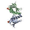
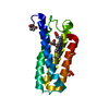
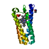
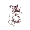
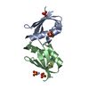
| Title | Crystal structure of cisplatin bound to a human copper chaperone (dimer) - new refinement | ||||||
 Components Components | Copper transport protein ATOX1 | ||||||
 Keywords Keywords | METAL TRANSPORT / Re-refinement of 3iwx / cisplatin / platinum / Metal-binding | ||||||
| Function / homology |  Function and homology information Function and homology informationmetallochaperone activity / Ion influx/efflux at host-pathogen interface / copper-dependent protein binding / copper chaperone activity / copper ion export / copper ion transport / Detoxification of Reactive Oxygen Species / cuprous ion binding / intracellular copper ion homeostasis / ATPase binding ...metallochaperone activity / Ion influx/efflux at host-pathogen interface / copper-dependent protein binding / copper chaperone activity / copper ion export / copper ion transport / Detoxification of Reactive Oxygen Species / cuprous ion binding / intracellular copper ion homeostasis / ATPase binding / response to oxidative stress / copper ion binding / negative regulation of apoptotic process / cytosol Similarity search - Function | ||||||
| Biological species |  Homo sapiens (human) Homo sapiens (human) | ||||||
| Method |  X-RAY DIFFRACTION / SYNCHROTRON / FOURIER SYNTHESIS / Resolution: 2.14 Å X-RAY DIFFRACTION / SYNCHROTRON / FOURIER SYNTHESIS / Resolution: 2.14 Å | ||||||
 Authors Authors | Shabalin, I.G. / Dauter, Z. / Jaskolski, M. / Minor, W. / Wlodawer, A. | ||||||
| Funding support |  United States, 1items United States, 1items
| ||||||
 Citation Citation | Journal: Acta Crystallogr.,Sect.D / Year: 2015 Title: Crystallography and chemistry should always go together: a cautionary tale of protein complexes with cisplatin and carboplatin. Authors: Shabalin, I. / Dauter, Z. / Jaskolski, M. / Minor, W. / Wlodawer, A. #1: Journal: J.Am.Chem.Soc. / Year: 2009Title: Crystal structures of cisplatin bound to a human copper chaperone. Authors: Boal, A.K. / Rosenzweig, A.C. | ||||||
| History |
|
- Structure visualization
| Structure viewer | Molecule: MolmilJmol/JSmol |
|---|
- Downloads & links
-Download
| PDBx/mmCIF format | 4yea.cif.gz | 70.4 KB | Display | PDBx/mmCIF format |
|---|---|---|---|---|
| PDB format | pdb4yea.ent.gz | 51.6 KB | Display | PDB format |
| PDBx/mmJSON format | 4yea.json.gz | Tree view | PDBx/mmJSON format | |
| Others |  Other downloads Other downloads |
-Validation report
| Arichive directory | https://data.pdbj.org/pub/pdb/validation_reports/ye/4yeaftp://data.pdbj.org/pub/pdb/validation_reports/ye/4yea | HTTPS FTP |
|---|
-Related structure data
| Related structure data | 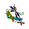 4ydxC  4yemC 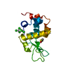 4yenC 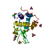 4yeoC  3iwxS S: Starting model for refinement C: citing same article ( |
|---|---|
| Similar structure data |


-Links
 PDBj
PDBj


- Assembly
| Deposited unit | 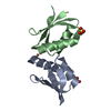
| ||||||||
|---|---|---|---|---|---|---|---|---|---|
| 1 |
| ||||||||
| Unit cell |
|
-Components
| #1: Protein | Mass: 7281.451 Da / Num. of mol.: 2 Source method: isolated from a genetically manipulated source Source: (gene. exp.) Homo sapiens (human) / Gene: ATOX1, HAH1 / Plasmid: pET21b / Production host:  #2: Chemical | ChemComp-CU / |   Mass: 63.546 Da / Num. of mol.: 1 / Source method: obtained synthetically / Formula: Cu Mass: 63.546 Da / Num. of mol.: 1 / Source method: obtained synthetically / Formula: Cu#3: Chemical |   Mass: 96.063 Da / Num. of mol.: 3 / Source method: obtained synthetically / Formula: SO4 Mass: 96.063 Da / Num. of mol.: 3 / Source method: obtained synthetically / Formula: SO4#4: Water | ChemComp-HOH / |  Mass: 18.015 Da / Num. of mol.: 111 / Source method: isolated from a natural source / Formula: H2O Mass: 18.015 Da / Num. of mol.: 111 / Source method: isolated from a natural source / Formula: H2O |
|---|
-Experimental details
-Experiment
| Experiment | Method: X-RAY DIFFRACTION / Number of used crystals: 1 |
|---|
- Sample preparation
| Crystal | Density Matthews: 3.24 Å3/Da / Density % sol: 62.06 % |
|---|---|
| Crystal grow | Temperature: 294 K / Method: vapor diffusion, hanging drop / pH: 6 Details: 1.5 M LITHIUM SULFATE, 0.1M MES, 50 MM NACL, PH 6, VAPOR DIFFUSION, HANGING DROP, TEMPERATURE 294K |
-Data collection
| Diffraction | Mean temperature: 113 K |
|---|---|
| Diffraction source | Source: SYNCHROTRON / Site: APS / Beamline: 21-ID-F / Wavelength: 0.97872 Å |
| Detector | Type: MARRESEARCH / Detector: CCD / Date: Apr 1, 2009 |
| Radiation | Protocol: SINGLE WAVELENGTH / Monochromatic (M) / Laue (L): M / Scattering type: x-ray |
| Radiation wavelength | Wavelength: 0.97872 Å / Relative weight: 1 |
| Reflection | Resolution: 2.14→50 Å / Num. obs: 10562 / % possible obs: 99.9 % / Redundancy: 5.6 % / Biso Wilson estimate: 33.3 Å2 / Rmerge(I) obs: 0.078 / Rsym value: 0.078 / Net I/σ(I): 23.4 |
| Reflection shell | Resolution: 2.14→2.18 Å / Redundancy: 5.5 % / Rmerge(I) obs: 0.401 / Mean I/σ(I) obs: 3.5 / % possible all: 100 |
- Processing
| Software |
| |||||||||||||||||||||||||||||||||||||||||||||||||||||||||||||||||||||||||||||||||||||||||||||||||||||||||||||||||||||||||||||||||||||||||||||||||||||||||||||||||||||||||||||||||||||||||||||||||||||||||||||||||||||||||||||||||||||||||||||||||||||||||||||||||||||||||||||||||||||||||||||||||||||||||||||||||||||||||||||||||||||
|---|---|---|---|---|---|---|---|---|---|---|---|---|---|---|---|---|---|---|---|---|---|---|---|---|---|---|---|---|---|---|---|---|---|---|---|---|---|---|---|---|---|---|---|---|---|---|---|---|---|---|---|---|---|---|---|---|---|---|---|---|---|---|---|---|---|---|---|---|---|---|---|---|---|---|---|---|---|---|---|---|---|---|---|---|---|---|---|---|---|---|---|---|---|---|---|---|---|---|---|---|---|---|---|---|---|---|---|---|---|---|---|---|---|---|---|---|---|---|---|---|---|---|---|---|---|---|---|---|---|---|---|---|---|---|---|---|---|---|---|---|---|---|---|---|---|---|---|---|---|---|---|---|---|---|---|---|---|---|---|---|---|---|---|---|---|---|---|---|---|---|---|---|---|---|---|---|---|---|---|---|---|---|---|---|---|---|---|---|---|---|---|---|---|---|---|---|---|---|---|---|---|---|---|---|---|---|---|---|---|---|---|---|---|---|---|---|---|---|---|---|---|---|---|---|---|---|---|---|---|---|---|---|---|---|---|---|---|---|---|---|---|---|---|---|---|---|---|---|---|---|---|---|---|---|---|---|---|---|---|---|---|---|---|---|---|---|---|---|---|---|---|---|---|---|---|---|---|---|---|---|---|---|---|---|---|---|---|---|---|---|---|---|---|---|---|---|---|---|---|---|---|---|---|---|---|---|---|---|---|---|---|---|---|---|---|---|---|---|---|---|---|---|---|---|---|---|
| Refinement | Method to determine structure: FOURIER SYNTHESIS Starting model: 3iwx Resolution: 2.14→25.63 Å / Cor.coef. Fo:Fc: 0.965 / Cor.coef. Fo:Fc free: 0.952 / WRfactor Rfree: 0.2055 / WRfactor Rwork: 0.162 / FOM work R set: 0.8799 / SU B: 7.506 / SU ML: 0.099 / SU R Cruickshank DPI: 0.1601 / SU Rfree: 0.1453 / Cross valid method: THROUGHOUT / σ(F): 0 / ESU R: 0.16 / ESU R Free: 0.145 / Stereochemistry target values: MAXIMUM LIKELIHOOD Details: U VALUES : WITH TLS ADDED HYDROGENS HAVE BEEN ADDED IN THE RIDING POSITIONS THIS DEPOSIT RESULTED FROM AN ANALYSIS OF A NUMBER OF PDB ENTRIES THAT CONTAIN CISPLATIN OR CARBOPLATIN IN COMPLEX ...Details: U VALUES : WITH TLS ADDED HYDROGENS HAVE BEEN ADDED IN THE RIDING POSITIONS THIS DEPOSIT RESULTED FROM AN ANALYSIS OF A NUMBER OF PDB ENTRIES THAT CONTAIN CISPLATIN OR CARBOPLATIN IN COMPLEX WITH PROTEINS, CONDUCTED IN THE SPIRIT OF THE TERWILLIGER-BRICOGNE MOTTO ADVOCATING CONTINUOUS IMPROVEMENT OF THE MACROMOLECULAR MODELS IN THE PDB (ACTA CRYST. D70, 2533, 2014). THE STRUCTURE FACTORS AND COORDINATES, ORIGINALLY DEPOSITED AS 3IWX (BOAL, A. K. & ROSENZWEIG, A. C. 2009. J. AM. CHEM. SOC. 131, 14196-14197), WERE USED AS THE STARTING POINT FOR AN INDEPENDENT RE-REFINEMENT. THE NEW MODEL INCLUDES SOME REINTERPRETATION OF THE LIGANDS, HAS LOWER R FACTORS, AND IMPROVED STATISTICS DESCRIBING THE AGREEMENT WITH THE EXPERIMENTAL DATA.
| |||||||||||||||||||||||||||||||||||||||||||||||||||||||||||||||||||||||||||||||||||||||||||||||||||||||||||||||||||||||||||||||||||||||||||||||||||||||||||||||||||||||||||||||||||||||||||||||||||||||||||||||||||||||||||||||||||||||||||||||||||||||||||||||||||||||||||||||||||||||||||||||||||||||||||||||||||||||||||||||||||||
| Solvent computation | Ion probe radii: 0.8 Å / Shrinkage radii: 0.8 Å / VDW probe radii: 1.2 Å / Solvent model: MASK | |||||||||||||||||||||||||||||||||||||||||||||||||||||||||||||||||||||||||||||||||||||||||||||||||||||||||||||||||||||||||||||||||||||||||||||||||||||||||||||||||||||||||||||||||||||||||||||||||||||||||||||||||||||||||||||||||||||||||||||||||||||||||||||||||||||||||||||||||||||||||||||||||||||||||||||||||||||||||||||||||||||
| Displacement parameters | Biso max: 69.1 Å2 / Biso mean: 36.165 Å2 / Biso min: 23.58 Å2
| |||||||||||||||||||||||||||||||||||||||||||||||||||||||||||||||||||||||||||||||||||||||||||||||||||||||||||||||||||||||||||||||||||||||||||||||||||||||||||||||||||||||||||||||||||||||||||||||||||||||||||||||||||||||||||||||||||||||||||||||||||||||||||||||||||||||||||||||||||||||||||||||||||||||||||||||||||||||||||||||||||||
| Refinement step | Cycle: final / Resolution: 2.14→25.63 Å
| |||||||||||||||||||||||||||||||||||||||||||||||||||||||||||||||||||||||||||||||||||||||||||||||||||||||||||||||||||||||||||||||||||||||||||||||||||||||||||||||||||||||||||||||||||||||||||||||||||||||||||||||||||||||||||||||||||||||||||||||||||||||||||||||||||||||||||||||||||||||||||||||||||||||||||||||||||||||||||||||||||||
| Refine LS restraints |
| |||||||||||||||||||||||||||||||||||||||||||||||||||||||||||||||||||||||||||||||||||||||||||||||||||||||||||||||||||||||||||||||||||||||||||||||||||||||||||||||||||||||||||||||||||||||||||||||||||||||||||||||||||||||||||||||||||||||||||||||||||||||||||||||||||||||||||||||||||||||||||||||||||||||||||||||||||||||||||||||||||||
| LS refinement shell | Resolution: 2.143→2.198 Å / Total num. of bins used: 20
| |||||||||||||||||||||||||||||||||||||||||||||||||||||||||||||||||||||||||||||||||||||||||||||||||||||||||||||||||||||||||||||||||||||||||||||||||||||||||||||||||||||||||||||||||||||||||||||||||||||||||||||||||||||||||||||||||||||||||||||||||||||||||||||||||||||||||||||||||||||||||||||||||||||||||||||||||||||||||||||||||||||
| Refinement TLS params. | Method: refined / Refine-ID: X-RAY DIFFRACTION
| |||||||||||||||||||||||||||||||||||||||||||||||||||||||||||||||||||||||||||||||||||||||||||||||||||||||||||||||||||||||||||||||||||||||||||||||||||||||||||||||||||||||||||||||||||||||||||||||||||||||||||||||||||||||||||||||||||||||||||||||||||||||||||||||||||||||||||||||||||||||||||||||||||||||||||||||||||||||||||||||||||||
| Refinement TLS group |
|
