[English] 日本語
Yorodumi
- PDB-3iwx: Crystal structure of cisplatin bound to a human copper chaperone ... -

+
Open data


ID or keywords:

Loading...

-
Basic information

Entry
Database: PDB / ID: 3iwx
Title
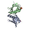
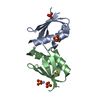
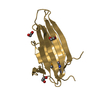
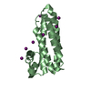
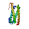
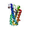
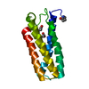
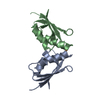
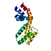
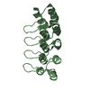
Crystal structure of cisplatin bound to a human copper chaperone (dimer)
ComponentsCopper transport protein ATOX1
KeywordsMETAL TRANSPORT / beta-alpha-beta-beta-alpha-beta / transport protein / cisplatin / platinum / Chaperone / Copper transport / Ion transport / Metal-binding / Transport
Function / homology
Function and homology information


metallochaperone activity / Ion influx/efflux at host-pathogen interface / copper-dependent protein binding / copper chaperone activity / copper ion transport / Detoxification of Reactive Oxygen Species / cuprous ion binding / intracellular copper ion homeostasis / response to oxidative stress / copper ion binding / cytosol
Similarity search - Function
Heavy-metal-associated, conserved site / Heavy-metal-associated domain. / Heavy-metal-associated domain / Heavy metal-associated domain superfamily / Heavy-metal-associated domain profile. / Heavy metal-associated domain, HMA / Alpha-Beta Plaits - #100 / Alpha-Beta Plaits / 2-Layer Sandwich / Alpha Beta
Similarity search - Domain/homology
Cisplatin / Copper transport protein ATOX1
Similarity search - Component
Biological speciesHomo sapiens (human)
MethodX-RAY DIFFRACTION / SYNCHROTRON / MOLECULAR REPLACEMENT / Resolution: 2.14 Å
AuthorsBoal, A.K. / Rosenzweig, A.C.
CitationJournal: J.Am.Chem.Soc. / Year: 2009
Title: Crystal structures of cisplatin bound to a human copper chaperone.
Authors: Boal, A.K. / Rosenzweig, A.C.
History
DepositionSep 3, 2009Deposition site: RCSB / Processing site: RCSB
Revision 1.0Sep 22, 2009Provider: repository / Type: Initial release
Revision 1.1Jul 13, 2011Group: Advisory / Refinement description / Version format compliance
Revision 1.2Feb 21, 2024Group: Data collection / Database references / Derived calculations
Category: chem_comp_atom / chem_comp_bond ...chem_comp_atom / chem_comp_bond / database_2 / pdbx_struct_conn_angle / struct_conn / struct_site
Item: _database_2.pdbx_DOI / _database_2.pdbx_database_accession ..._database_2.pdbx_DOI / _database_2.pdbx_database_accession / _pdbx_struct_conn_angle.ptnr1_auth_asym_id / _pdbx_struct_conn_angle.ptnr1_auth_seq_id / _pdbx_struct_conn_angle.ptnr1_label_asym_id / _pdbx_struct_conn_angle.ptnr1_label_seq_id / _pdbx_struct_conn_angle.ptnr3_auth_asym_id / _pdbx_struct_conn_angle.ptnr3_auth_seq_id / _pdbx_struct_conn_angle.ptnr3_label_asym_id / _pdbx_struct_conn_angle.ptnr3_label_seq_id / _pdbx_struct_conn_angle.value / _struct_conn.pdbx_dist_value / _struct_conn.ptnr1_auth_asym_id / _struct_conn.ptnr1_auth_comp_id / _struct_conn.ptnr1_auth_seq_id / _struct_conn.ptnr1_label_asym_id / _struct_conn.ptnr1_label_atom_id / _struct_conn.ptnr1_label_comp_id / _struct_conn.ptnr1_label_seq_id / _struct_conn.ptnr2_auth_asym_id / _struct_conn.ptnr2_auth_comp_id / _struct_conn.ptnr2_auth_seq_id / _struct_conn.ptnr2_label_asym_id / _struct_conn.ptnr2_label_atom_id / _struct_conn.ptnr2_label_comp_id / _struct_conn.ptnr2_label_seq_id / _struct_site.pdbx_auth_asym_id / _struct_site.pdbx_auth_comp_id / _struct_site.pdbx_auth_seq_id

-
Structure visualization

Structure viewerMolecule:
MolmilJmol/JSmol

Downloads & links

-
Assembly

Deposited unit
A: Copper transport protein ATOX1
B: Copper transport protein ATOX1
hetero molecules


Theoretical massNumber of molelcules
Total (without water)15,4146
Polymers14,8252
Non-polymers5884
Water1,892105
1


  • Idetical with deposited unit
  • defined by author&software
TypeNameSymmetry operationNumber
identity operation1_555x,y,z1
Buried area1740 Å2
ΔGint-58 kcal/mol
Surface area7070 Å2
MethodPISA
Unit cell
Length a, b, c (Å)78.289, 78.289, 54.335
Angle α, β, γ (deg.)90.00, 90.00, 120.00
Int Tables number170
Space group name H-MP65

-
Components

#1: Protein Copper transport protein ATOX1 / Metal transport protein ATX1


Mass: 7412.646 Da / Num. of mol.: 2
Source method: isolated from a genetically manipulated source
Source: (gene. exp.) Homo sapiens (human) / Gene: ATOX1, HAH1 / Plasmid: pET21b / Production host: Escherichia coli (E. coli) / Strain (production host): BL21(DE3) / References: UniProt: O00244
#2: Chemical ChemComp-CPT / Cisplatin / diammine(dichloro)platinum


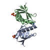
Mass: 300.045 Da / Num. of mol.: 1 / Source method: obtained synthetically / Formula: Cl2H6N2Pt / Comment: medication, chemotherapy*YM
#3: Chemical ChemComp-SO4 / SULFATE ION


Mass: 96.063 Da / Num. of mol.: 3 / Source method: obtained synthetically / Formula: SO4
#4: Water ChemComp-HOH / water


Mass: 18.015 Da / Num. of mol.: 105 / Source method: isolated from a natural source / Formula: H2O

-
Experimental details

-
Experiment

ExperimentMethod: X-RAY DIFFRACTION / Number of used crystals: 1

-
Sample preparation

CrystalDensity Matthews: 3.24 Å3/Da / Density % sol: 62.06 %
Crystal growTemperature: 294 K / Method: vapor diffusion, hanging drop / pH: 6
Details: 1.5 M lithium sulfate, 0.1M MES, 50 mM NaCl, pH 6, VAPOR DIFFUSION, HANGING DROP, temperature 294K

-
Data collection

DiffractionMean temperature: 113 K
Diffraction sourceSource: SYNCHROTRON / Site: APS / Beamline: 21-ID-F / Wavelength: 0.97872 Å
DetectorType: MARRESEARCH / Detector: CCD / Date: Apr 1, 2009
RadiationProtocol: SINGLE WAVELENGTH / Monochromatic (M) / Laue (L): M / Scattering type: x-ray
Radiation wavelengthWavelength: 0.97872 Å / Relative weight: 1
ReflectionResolution: 2.14→50 Å / Num. obs: 10562

-
Processing

Software
NameVersionClassificationNB
REFMAC5.5.0088refinement
PDB_EXTRACT3.005data extraction
RefinementMethod to determine structure: MOLECULAR REPLACEMENT / Resolution: 2.14→28.76 Å / Cor.coef. Fo:Fc: 0.953 / Cor.coef. Fo:Fc free: 0.934 / Occupancy max: 1 / Occupancy min: 0.4 / SU B: 9.17 / SU ML: 0.114 / TLS residual ADP flag: LIKELY RESIDUAL / Cross valid method: THROUGHOUT / σ(F): 0 / ESU R: 0.183 / ESU R Free: 0.167 / Stereochemistry target values: MAXIMUM LIKELIHOOD
Details: HYDROGENS HAVE BEEN ADDED IN THE RIDING POSITIONS. U VALUES: RESIDUAL ONLY
RfactorNum. reflection% reflectionSelection details
Rfree0.228 504 4.8 %RANDOM
Rwork0.186 ---
obs0.188 10536 99.91 %-
Solvent computationIon probe radii: 0.8 Å / Shrinkage radii: 0.8 Å / VDW probe radii: 1.2 Å / Solvent model: MASK
Displacement parametersBiso max: 102.23 Å2 / Biso mean: 24.288 Å2 / Biso min: 13.16 Å2
Baniso -1Baniso -2Baniso -3
1-0.06 Å20.03 Å20 Å2
2--0.06 Å20 Å2
3----0.09 Å2
Refinement stepCycle: LAST / Resolution: 2.14→28.76 Å
ProteinNucleic acidLigandSolventTotal
Num. atoms1011 0 18 105 1134
Refine LS restraints
Refine-IDTypeDev idealDev ideal targetNumber
X-RAY DIFFRACTIONr_bond_refined_d0.0090.0221034
X-RAY DIFFRACTIONr_angle_refined_deg1.0922.011389
X-RAY DIFFRACTIONr_dihedral_angle_1_deg5.6065132
X-RAY DIFFRACTIONr_dihedral_angle_2_deg37.66225.88234
X-RAY DIFFRACTIONr_dihedral_angle_3_deg12.56715201
X-RAY DIFFRACTIONr_dihedral_angle_4_deg22.208152
X-RAY DIFFRACTIONr_chiral_restr0.0720.2164
X-RAY DIFFRACTIONr_gen_planes_refined0.0040.02708
X-RAY DIFFRACTIONr_mcbond_it0.4461.5661
X-RAY DIFFRACTIONr_mcangle_it0.89821062
X-RAY DIFFRACTIONr_scbond_it1.6243373
X-RAY DIFFRACTIONr_scangle_it2.5974.5327
LS refinement shellResolution: 2.142→2.198 Å / Total num. of bins used: 20
RfactorNum. reflection% reflection
Rfree0.266 37 -
Rwork0.206 727 -
all-764 -
obs--100 %
Refinement TLS params.

Method: refined / Refine-ID: X-RAY DIFFRACTION

IDL112)L122)L132)L222)L232)L332)S11 (Å °)S12 (Å °)S13 (Å °)S21 (Å °)S22 (Å °)S23 (Å °)S31 (Å °)S32 (Å °)S33 (Å °)T112)T122)T132)T222)T232)T332)Origin x (Å)Origin y (Å)Origin z (Å)
13.5144-0.53510.36094.6825-0.30494.1899-0.1489-0.02960.11030.01410.0555-0.0473-0.1814-0.07650.09340.03580.0113-0.00590.00670.01270.09721.8823-1.98054.503
22.12080.35671.31233.16261.09114.4005-0.03430.05310.0626-0.00430.1083-0.0421-0.02650.1345-0.0740.08520.00050.03250.04690.01280.030939.3026-16.54923.1873
Refinement TLS group
IDRefine-IDRefine TLS-IDAuth asym-IDAuth seq-ID
1X-RAY DIFFRACTION1A2 - 68
2X-RAY DIFFRACTION1A69 - 71
3X-RAY DIFFRACTION1A72 - 113
4X-RAY DIFFRACTION2B2 - 68
5X-RAY DIFFRACTION2B70 - 71
6X-RAY DIFFRACTION2B72 - 134

+
About Yorodumi

-
News

-
Feb 9, 2022. New format data for meta-information of EMDB entries

New format data for meta-information of EMDB entries

  • Version 3 of the EMDB header file is now the official format.
  • The previous official version 1.9 will be removed from the archive.

Related info.:EMDB header

External links:wwPDB to switch to version 3 of the EMDB data model

-
Aug 12, 2020. Covid-19 info

Covid-19 info

URL: https://pdbjlvh1.pdbj.org/emnavi/covid19.php

New page: Covid-19 featured information page in EM Navigator.

Related info.:Covid-19 info / Mar 5, 2020. Novel coronavirus structure data

+
Mar 5, 2020. Novel coronavirus structure data

Novel coronavirus structure data

Related info.:Yorodumi Speices / Aug 12, 2020. Covid-19 info

External links:COVID-19 featured content - PDBj / Molecule of the Month (242):Coronavirus Proteases

+
Jan 31, 2019. EMDB accession codes are about to change! (news from PDBe EMDB page)

EMDB accession codes are about to change! (news from PDBe EMDB page)

  • The allocation of 4 digits for EMDB accession codes will soon come to an end. Whilst these codes will remain in use, new EMDB accession codes will include an additional digit and will expand incrementally as the available range of codes is exhausted. The current 4-digit format prefixed with “EMD-” (i.e. EMD-XXXX) will advance to a 5-digit format (i.e. EMD-XXXXX), and so on. It is currently estimated that the 4-digit codes will be depleted around Spring 2019, at which point the 5-digit format will come into force.
  • The EM Navigator/Yorodumi systems omit the EMD- prefix.

Related info.:Q: What is EMD? / ID/Accession-code notation in Yorodumi/EM Navigator

External links:EMDB Accession Codes are Changing Soon! / Contact to PDBj

+
Jul 12, 2017. Major update of PDB

Major update of PDB

  • wwPDB released updated PDB data conforming to the new PDBx/mmCIF dictionary.
  • This is a major update changing the version number from 4 to 5, and with Remediation, in which all the entries are updated.
  • In this update, many items about electron microscopy experimental information are reorganized (e.g. em_software).
  • Now, EM Navigator and Yorodumi are based on the updated data.

External links:wwPDB Remediation / Enriched Model Files Conforming to OneDep Data Standards Now Available in the PDB FTP Archive

-
Yorodumi

Thousand views of thousand structures

  • Yorodumi is a browser for structure data from EMDB, PDB, SASBDB, etc.
  • This page is also the successor to EM Navigator detail page, and also detail information page/front-end page for Omokage search.
  • The word "yorodu" (or yorozu) is an old Japanese word meaning "ten thousand". "mi" (miru) is to see.

Related info.:EMDB / PDB / SASBDB / Comparison of 3 databanks / Yorodumi Search / Aug 31, 2016. New EM Navigator & Yorodumi / Yorodumi Papers / Jmol/JSmol / Function and homology information / Changes in new EM Navigator and Yorodumi

Read more