 Movie
Movie Controller

[English] 日本語
 Yorodumi
- PDB-2cj8: Crystal Structure of a Cell Wall Invertase Inhibitor from Tobacco... -
+ Open data

- Basic information
| Entry | Database: PDB / ID: 2cj8 | ||||||
|---|---|---|---|---|---|---|---|
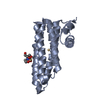
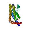
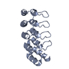
| Title | Crystal Structure of a Cell Wall Invertase Inhibitor from Tobacco (pH 9.5) | ||||||
 Components Components | INVERTASE INHIBITOR | ||||||
 Keywords Keywords | INHIBITOR / PROTEIN BINDING / FOUR-HELIX BUNDLE / HELICAL HAIRPIN | ||||||
| Function / homology |  Function and homology information Function and homology informationplant-type cell wall modification / plant-type cell wall / enzyme inhibitor activity Similarity search - Function | ||||||
| Biological species |  | ||||||
| Method |  X-RAY DIFFRACTION / SYNCHROTRON / MOLECULAR REPLACEMENT / Resolution: 2.38 Å X-RAY DIFFRACTION / SYNCHROTRON / MOLECULAR REPLACEMENT / Resolution: 2.38 Å | ||||||
 Authors Authors | Hothorn, M. / Scheffzek, K. | ||||||
 Citation Citation | Journal: Acta Crystallogr.,Sect.D / Year: 2006 Title: Multiple Crystal Forms of the Cell-Wall Invertase Inhibitor from Tobacco Support High Conformational Rigidity Over a Broad Ph-Range Authors: Hothorn, M. / Scheffzek, K. | ||||||
| History |
|
- Structure visualization
| Structure viewer | Molecule: MolmilJmol/JSmol |
|---|
- Downloads & links
-Download
| PDBx/mmCIF format | 2cj8.cif.gz | 66.6 KB | Display | PDBx/mmCIF format |
|---|---|---|---|---|
| PDB format | pdb2cj8.ent.gz | 49.7 KB | Display | PDB format |
| PDBx/mmJSON format | 2cj8.json.gz | Tree view | PDBx/mmJSON format | |
| Others |  Other downloads Other downloads |
-Validation report
| Arichive directory | https://data.pdbj.org/pub/pdb/validation_reports/cj/2cj8ftp://data.pdbj.org/pub/pdb/validation_reports/cj/2cj8 | HTTPS FTP |
|---|
-Related structure data
| Related structure data |  2cj4C  2cj5C  2cj6C  2cj7C 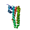 1rj1S S: Starting model for refinement C: citing same article ( |
|---|---|
| Similar structure data |


-Links
 PDBj
PDBj- Assembly
| Deposited unit | 
| ||||||||
|---|---|---|---|---|---|---|---|---|---|
| 1 | 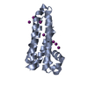
| ||||||||
| 2 | 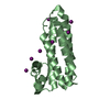
| ||||||||
| Unit cell |
|
-Components
| #1: Protein | Mass: 16031.401 Da / Num. of mol.: 2 Source method: isolated from a genetically manipulated source Source: (gene. exp.)  #2: Chemical | ChemComp-IOD /   Mass: 126.904 Da / Num. of mol.: 18 / Source method: obtained synthetically / Formula: I Mass: 126.904 Da / Num. of mol.: 18 / Source method: obtained synthetically / Formula: I#3: Water | ChemComp-HOH / |  Mass: 18.015 Da / Num. of mol.: 50 / Source method: isolated from a natural source / Formula: H2O Mass: 18.015 Da / Num. of mol.: 50 / Source method: isolated from a natural source / Formula: H2OHas protein modification | Y | |
|---|
-Experimental details
-Experiment
| Experiment | Method: X-RAY DIFFRACTION / Number of used crystals: 1 |
|---|
- Sample preparation
| Crystal | Density Matthews: 2.1 Å3/Da / Density % sol: 39.9 % |
|---|---|
| Crystal grow | pH: 9.5 / Details: 18 % PEG 4000, 0.1 M TRICINE PH 9.5, 0.2 M NAI |
-Data collection
| Diffraction | Mean temperature: 100 K |
|---|---|
| Diffraction source | Source: SYNCHROTRON / Site: EMBL/DESY, HAMBURG  / Beamline: BW7A / Wavelength: 0.976 / Beamline: BW7A / Wavelength: 0.976 |
| Detector | Type: MARRESEARCH / Detector: CCD / Date: Feb 26, 2003 |
| Radiation | Protocol: SINGLE WAVELENGTH / Monochromatic (M) / Laue (L): M / Scattering type: x-ray |
| Radiation wavelength | Wavelength: 0.976 Å / Relative weight: 1 |
| Reflection | Resolution: 2.38→19.21 Å / Num. obs: 11192 / % possible obs: 100 % / Observed criterion σ(I): -3 / Redundancy: 7.1 % / Rmerge(I) obs: 0.12 / Net I/σ(I): 13.6 |
| Reflection shell | Resolution: 2.38→2.58 Å / Redundancy: 7.2 % / Rmerge(I) obs: 0.45 / Mean I/σ(I) obs: 4.4 / % possible all: 99.7 |
- Processing
| Software |
| ||||||||||||||||||||||||||||||||||||||||||||||||||||||||||||||||||||||||||||||||||||||||||||||||||||||||||||||||||||||||||||||||||||||||||||||||||||||||||||||||||||||||||||||||||||||
|---|---|---|---|---|---|---|---|---|---|---|---|---|---|---|---|---|---|---|---|---|---|---|---|---|---|---|---|---|---|---|---|---|---|---|---|---|---|---|---|---|---|---|---|---|---|---|---|---|---|---|---|---|---|---|---|---|---|---|---|---|---|---|---|---|---|---|---|---|---|---|---|---|---|---|---|---|---|---|---|---|---|---|---|---|---|---|---|---|---|---|---|---|---|---|---|---|---|---|---|---|---|---|---|---|---|---|---|---|---|---|---|---|---|---|---|---|---|---|---|---|---|---|---|---|---|---|---|---|---|---|---|---|---|---|---|---|---|---|---|---|---|---|---|---|---|---|---|---|---|---|---|---|---|---|---|---|---|---|---|---|---|---|---|---|---|---|---|---|---|---|---|---|---|---|---|---|---|---|---|---|---|---|---|
| Refinement | Method to determine structure: MOLECULAR REPLACEMENT Starting model: PDB ENTRY 1RJ1 Resolution: 2.38→19.21 Å / Cor.coef. Fo:Fc: 0.929 / Cor.coef. Fo:Fc free: 0.859 / SU B: 14.218 / SU ML: 0.221 / TLS residual ADP flag: LIKELY RESIDUAL / Cross valid method: THROUGHOUT / ESU R: 0.536 / ESU R Free: 0.302 / Stereochemistry target values: MAXIMUM LIKELIHOOD / Details: HYDROGENS HAVE BEEN ADDED IN THE RIDING POSITIONS
| ||||||||||||||||||||||||||||||||||||||||||||||||||||||||||||||||||||||||||||||||||||||||||||||||||||||||||||||||||||||||||||||||||||||||||||||||||||||||||||||||||||||||||||||||||||||
| Solvent computation | Ion probe radii: 0.8 Å / Shrinkage radii: 0.8 Å / VDW probe radii: 1.2 Å / Solvent model: MASK | ||||||||||||||||||||||||||||||||||||||||||||||||||||||||||||||||||||||||||||||||||||||||||||||||||||||||||||||||||||||||||||||||||||||||||||||||||||||||||||||||||||||||||||||||||||||
| Displacement parameters | Biso mean: 37.44 Å2
| ||||||||||||||||||||||||||||||||||||||||||||||||||||||||||||||||||||||||||||||||||||||||||||||||||||||||||||||||||||||||||||||||||||||||||||||||||||||||||||||||||||||||||||||||||||||
| Refinement step | Cycle: LAST / Resolution: 2.38→19.21 Å
| ||||||||||||||||||||||||||||||||||||||||||||||||||||||||||||||||||||||||||||||||||||||||||||||||||||||||||||||||||||||||||||||||||||||||||||||||||||||||||||||||||||||||||||||||||||||
| Refine LS restraints |
|