Resolution: 1.9→25 Å / Cor.coef. Fo:Fc: 0.952 / Cor.coef. Fo:Fc free: 0.92 / SU B: 3.432 / SU ML: 0.102 / Cross valid method: THROUGHOUT / ESU R: 0.156 / ESU R Free: 0.149 / Stereochemistry target values: MAXIMUM LIKELIHOOD / Details: HYDROGENS HAVE BEEN ADDED IN THE RIDING POSITIONS.
Rfactor
Num. reflection
% reflection
Selection details
Rfree
0.227
1169
4.8 %
RANDOM
Rwork
0.176
-
-
-
obs
0.178
23178
99.9 %
-
Solvent computation
Ion probe radii: 0.8 Å / Shrinkage radii: 0.8 Å / VDW probe radii: 1.4 Å / Solvent model: BABINET MODEL WITH MASK
 Movie
Movie Controller


 Open data
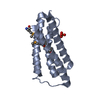
 Basic information
 Components
 Keywords
 Function and homology information
 X-RAY DIFFRACTION /
  Authors
 Citation
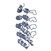
 Structure visualization
 Downloads & links
 Other downloads


 PDBj
PDBj

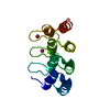
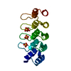
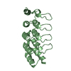
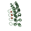
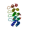
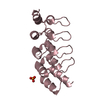
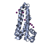
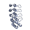
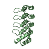
 Assembly

 Mass: 18.015 Da / Num. of mol.: 330 / Source method: isolated from a natural source / Formula: H2O
 Sample preparation
 / Beamline: X06SA / Wavelength: 0.9
  Processing