 Movie
Movie Controller

[English] 日本語
 Yorodumi
- PDB-1rj1: Crystal Structure of a Cell Wall Invertase Inhibitor from Tobacco -
+ Open data

- Basic information
| Entry | Database: PDB / ID: 1rj1 | ||||||
|---|---|---|---|---|---|---|---|
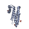
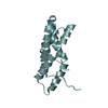
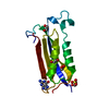
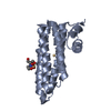
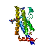
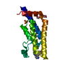
| Title | Crystal Structure of a Cell Wall Invertase Inhibitor from Tobacco | ||||||
 Components Components | invertase inhibitor | ||||||
 Keywords Keywords | PROTEIN BINDING / FOUR-HELIX BUNDLE / HELICAL HAIRPIN | ||||||
| Function / homology |  Function and homology information Function and homology informationplant-type cell wall modification / plant-type cell wall / enzyme inhibitor activity Similarity search - Function | ||||||
| Biological species |  | ||||||
| Method |  X-RAY DIFFRACTION / SYNCHROTRON / MOLECULAR REPLACEMENT / Resolution: 1.87 Å X-RAY DIFFRACTION / SYNCHROTRON / MOLECULAR REPLACEMENT / Resolution: 1.87 Å | ||||||
 Authors Authors | Hothorn, M. / D'Angelo, I. / Marquez, J.A. / Greiner, S. / Scheffzek, K. | ||||||
 Citation Citation | Journal: J.Mol.Biol. / Year: 2004 Title: The invertase inhibitor Nt-CIF from tobacco: a highly thermostable four-helix bundle with an unusual N-terminal extension Authors: Hothorn, M. / D'Angelo, I. / Marquez, J.A. / Greiner, S. / Scheffzek, K. | ||||||
| History |
|
- Structure visualization
| Structure viewer | Molecule: MolmilJmol/JSmol |
|---|
- Downloads & links
-Download
| PDBx/mmCIF format | 1rj1.cif.gz | 41.9 KB | Display | PDBx/mmCIF format |
|---|---|---|---|---|
| PDB format | pdb1rj1.ent.gz | 28.9 KB | Display | PDB format |
| PDBx/mmJSON format | 1rj1.json.gz | Tree view | PDBx/mmJSON format | |
| Others |  Other downloads Other downloads |
-Validation report
| Arichive directory | https://data.pdbj.org/pub/pdb/validation_reports/rj/1rj1ftp://data.pdbj.org/pub/pdb/validation_reports/rj/1rj1 | HTTPS FTP |
|---|
-Related structure data
| Related structure data |  1rj4SC S: Starting model for refinement C: citing same article ( |
|---|---|
| Similar structure data |
-Links
 PDBj
PDBj- Assembly
| Deposited unit | 
| ||||||||
|---|---|---|---|---|---|---|---|---|---|
| 1 |
| ||||||||
| Unit cell |
| ||||||||
| Details | The biological assembly is a monomer in solution |
-Components
| #1: Protein | Mass: 16088.454 Da / Num. of mol.: 1 Source method: isolated from a genetically manipulated source Source: (gene. exp.)  |
|---|---|
| #2: Water | ChemComp-HOH /  Mass: 18.015 Da / Num. of mol.: 121 / Source method: isolated from a natural source / Formula: H2O Mass: 18.015 Da / Num. of mol.: 121 / Source method: isolated from a natural source / Formula: H2O |
| Has protein modification | Y |
-Experimental details
-Experiment
| Experiment | Method: X-RAY DIFFRACTION / Number of used crystals: 2 |
|---|
- Sample preparation
| Crystal | Density Matthews: 2.62 Å3/Da / Density % sol: 52.68 % | ||||||||||||||||||||||||
|---|---|---|---|---|---|---|---|---|---|---|---|---|---|---|---|---|---|---|---|---|---|---|---|---|---|
| Crystal grow | Temperature: 293 K / Method: vapor diffusion, hanging drop / pH: 7 Details: 4.0M sodium formate, 0.1M bis-Tris, pH 7.0, VAPOR DIFFUSION, HANGING DROP, temperature 293K | ||||||||||||||||||||||||
| Crystal grow | *PLUS pH: 7 / Method: vapor diffusion, hanging drop / Details: Hothorn, M., (2003) Acta Cryst., D59, 2279. | ||||||||||||||||||||||||
| Components of the solutions | *PLUS
|
-Data collection
| Diffraction | Mean temperature: 100 K |
|---|---|
| Diffraction source | Source: SYNCHROTRON / Site: ESRF  / Beamline: ID14-4 / Wavelength: 0.9393 Å / Beamline: ID14-4 / Wavelength: 0.9393 Å |
| Detector | Type: ADSC QUANTUM 4 / Detector: CCD / Date: Jun 20, 2002 |
| Radiation | Protocol: SINGLE WAVELENGTH / Monochromatic (M) / Laue (L): M / Scattering type: x-ray |
| Radiation wavelength | Wavelength: 0.9393 Å / Relative weight: 1 |
| Reflection | Resolution: 1.87→19.25 Å / Num. all: 15311 / Num. obs: 15112 / % possible obs: 98.7 % / Observed criterion σ(F): -3 / Observed criterion σ(I): -3 / Redundancy: 8.3 % / Biso Wilson estimate: 17.8 Å2 / Rmerge(I) obs: 0.057 / Net I/σ(I): 23.84 |
| Reflection shell | Resolution: 1.87→1.99 Å / Redundancy: 5.3 % / Rmerge(I) obs: 0.127 / Mean I/σ(I) obs: 8.92 / Num. unique all: 2210 / % possible all: 93.1 |
| Reflection | *PLUS Num. obs: 15167 / % possible obs: 97.8 % |
| Reflection shell | *PLUS Lowest resolution: 2 Å / % possible obs: 88.2 % / Num. unique obs: 2455 |
- Processing
| Software |
| ||||||||||||||||||||||||||||||||||||
|---|---|---|---|---|---|---|---|---|---|---|---|---|---|---|---|---|---|---|---|---|---|---|---|---|---|---|---|---|---|---|---|---|---|---|---|---|---|
| Refinement | Method to determine structure: MOLECULAR REPLACEMENT Starting model: PDB 1RJ4 Resolution: 1.87→19.25 Å / Rfactor Rfree error: 0.009 / Data cutoff high absF: 2543413.23 / Data cutoff low absF: 0 / Isotropic thermal model: RESTRAINED / Cross valid method: THROUGHOUT / σ(F): 0 / Stereochemistry target values: Engh & Huber
| ||||||||||||||||||||||||||||||||||||
| Solvent computation | Solvent model: FLAT MODEL / Bsol: 45.2858 Å2 / ksol: 0.385833 e/Å3 | ||||||||||||||||||||||||||||||||||||
| Displacement parameters | Biso mean: 21 Å2
| ||||||||||||||||||||||||||||||||||||
| Refine analyze |
| ||||||||||||||||||||||||||||||||||||
| Refinement step | Cycle: LAST / Resolution: 1.87→19.25 Å
| ||||||||||||||||||||||||||||||||||||
| Refine LS restraints |
| ||||||||||||||||||||||||||||||||||||
| LS refinement shell | Resolution: 1.87→1.99 Å / Rfactor Rfree error: 0.023 / Total num. of bins used: 6
| ||||||||||||||||||||||||||||||||||||
| Xplor file |
| ||||||||||||||||||||||||||||||||||||
| Refine LS restraints | *PLUS
|
