- PDB-4qot: Crystal structure of human copper chaperone bound to the platinum ion -
+
Open data
ID or keywords:
Loading...
-
Basic information
Entry
Database: PDB / ID: 4qot
Title
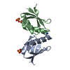
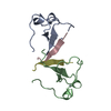
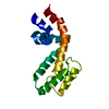
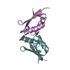
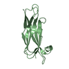
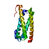
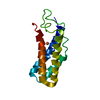
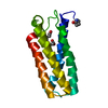
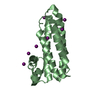
Crystal structure of human copper chaperone bound to the platinum ion
Components
Copper transport protein ATOX1
Keywords
CHAPERONE / Metal-binding / Metal transport / platinum / oxaliplatin
Function / homology
Function and homology information
metallochaperone activity / Ion influx/efflux at host-pathogen interface / copper-dependent protein binding / copper chaperone activity / copper ion export / copper ion transport / Detoxification of Reactive Oxygen Species / cuprous ion binding / intracellular copper ion homeostasis / ATPase binding ...metallochaperone activity / Ion influx/efflux at host-pathogen interface / copper-dependent protein binding / copper chaperone activity / copper ion export / copper ion transport / Detoxification of Reactive Oxygen Species / cuprous ion binding / intracellular copper ion homeostasis / ATPase binding / response to oxidative stress / copper ion binding / negative regulation of apoptotic process / cytosol Similarity search - Function
Resolution: 2.2→42.49 Å / SU ML: 0.28 / σ(F): 1.35 / Phase error: 26.01 / Stereochemistry target values: ML
Rfactor
Num. reflection
% reflection
Rfree
0.239
465
4.82 %
Rwork
0.188
-
-
obs
0.19
9647
99.3 %
all
-
9697
-
Solvent computation
Shrinkage radii: 0.9 Å / VDW probe radii: 1.11 Å / Solvent model: FLAT BULK SOLVENT MODEL
Refinement step
Cycle: LAST / Resolution: 2.2→42.49 Å
Protein
Nucleic acid
Ligand
Solvent
Total
Num. atoms
1012
0
27
48
1087
Refine LS restraints
Refine-ID
Type
Dev ideal
Number
X-RAY DIFFRACTION
f_bond_d
0.008
1077
X-RAY DIFFRACTION
f_angle_d
1.063
1444
X-RAY DIFFRACTION
f_dihedral_angle_d
13.544
400
X-RAY DIFFRACTION
f_chiral_restr
0.076
167
X-RAY DIFFRACTION
f_plane_restr
0.008
176
LS refinement shell
Resolution (Å)
Rfactor Rfree
Num. reflection Rfree
Rfactor Rwork
Num. reflection Rwork
Refine-ID
% reflection obs (%)
2.2012-2.5197
0.287
156
0.2429
3026
X-RAY DIFFRACTION
99
2.5197-3.1745
0.2728
169
0.2236
3050
X-RAY DIFFRACTION
100
3.1745-42.4961
0.206
140
0.1604
3106
X-RAY DIFFRACTION
99
Refinement TLS params.
Method: refined / Refine-ID: X-RAY DIFFRACTION
ID
L11 (°2)
L12 (°2)
L13 (°2)
L22 (°2)
L23 (°2)
L33 (°2)
S11 (Å °)
S12 (Å °)
S13 (Å °)
S21 (Å °)
S22 (Å °)
S23 (Å °)
S31 (Å °)
S32 (Å °)
S33 (Å °)
T11 (Å2)
T12 (Å2)
T13 (Å2)
T22 (Å2)
T23 (Å2)
T33 (Å2)
Origin x (Å)
Origin y (Å)
Origin z (Å)
1
0.0283
0.1659
0.2118
0.9681
1.2453
1.6118
0.1033
0.1397
-0.0239
-0.1434
-0.0912
0.0525
0.1025
-0.0179
-0.0067
0.3115
-0.0461
-0.0388
0.2318
0.063
0.2162
24.2328
151.454
-34.9802
2
1.4635
0.0344
-0.3366
3.4321
1.5297
2.4523
0.0116
-0.0879
0.1649
0.0072
0.1063
-0.0913
-0.2668
0.2624
-0.1236
0.4844
-0.1155
-0.1849
0.286
-0.1165
0.3927
43.2217
159.6953
-36.0526
Refinement TLS group
ID
Refine-ID
Refine TLS-ID
Selection details
1
X-RAY DIFFRACTION
1
(chainAandresseq15:15) or (chainAandresseq12:12) or (chainBandresseq15:15) or (chainBandresseq12:12) or (chainAandresseq101:101)
2
X-RAY DIFFRACTION
2
(chainBandresseq5:5andaltloc 'B') or (chainBandresseq101:101) or (chainBandresseq104:104) or (chainBandresseq41:41andaltloc 'A') or (chainBandresseq3:3andaltloc 'A')
+
About Yorodumi
-
News
-
Feb 9, 2022. New format data for meta-information of EMDB entries
New format data for meta-information of EMDB entries
Version 3 of the EMDB header file is now the official format.
The previous official version 1.9 will be removed from the archive.
In the structure databanks used in Yorodumi, some data are registered as the other names, "COVID-19 virus" and "2019-nCoV". Here are the details of the virus and the list of structure data.
Jan 31, 2019. EMDB accession codes are about to change! (news from PDBe EMDB page)
EMDB accession codes are about to change! (news from PDBe EMDB page)
The allocation of 4 digits for EMDB accession codes will soon come to an end. Whilst these codes will remain in use, new EMDB accession codes will include an additional digit and will expand incrementally as the available range of codes is exhausted. The current 4-digit format prefixed with “EMD-” (i.e. EMD-XXXX) will advance to a 5-digit format (i.e. EMD-XXXXX), and so on. It is currently estimated that the 4-digit codes will be depleted around Spring 2019, at which point the 5-digit format will come into force.
The EM Navigator/Yorodumi systems omit the EMD- prefix.
Related info.:Q: What is EMD? / ID/Accession-code notation in Yorodumi/EM Navigator
Yorodumi is a browser for structure data from EMDB, PDB, SASBDB, etc.
This page is also the successor to EM Navigator detail page, and also detail information page/front-end page for Omokage search.
The word "yorodu" (or yorozu) is an old Japanese word meaning "ten thousand". "mi" (miru) is to see.
Related info.:EMDB / PDB / SASBDB / Comparison of 3 databanks / Yorodumi Search / Aug 31, 2016. New EM Navigator & Yorodumi / Yorodumi Papers / Jmol/JSmol / Function and homology information / Changes in new EM Navigator and Yorodumi
 Movie
Movie Controller

 Yorodumi
 Open data
 Basic information
 Components
 Keywords
 Function and homology information
 Homo sapiens (human)
 X-RAY DIFFRACTION /
  Authors
 Citation
 Structure visualization
 Downloads & links
 Other downloads


 PDBj
PDBj


 Assembly
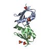


 Mass: 195.078 Da / Num. of mol.: 2 / Source method: obtained synthetically / Formula: Pt
 Mass: 96.063 Da / Num. of mol.: 5 / Source method: obtained synthetically / Formula: SO4
 Mass: 18.015 Da / Num. of mol.: 48 / Source method: isolated from a natural source / Formula: H2O
 Sample preparation
 / Beamline: ID29 / Wavelength: 1.071564 Å
 Processing