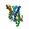
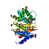
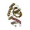
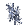
Entry Database : PDB / ID : 4xx2Title Ohr from Xylella fastidiosa in oxidized state Organic hydroperoxide resistance protein Keywords / / / / / Function / homology Function Domain/homology Component
/ / / / / / / / / / / / / / / / / / / / / Biological species Xylella fastidiosa (bacteria)Method / / / Resolution : 2.15 Å Authors Alegria, T.G.P. / Discola, K.F. / Cussiol, J.R.R. / Oliveira, M.A. / Netto, L.E.S. Funding support Organization Grant number Country Sao Paulo Research Foundation (FAPESP) 2008/07971-3
Journal : To Be Published Title : Ohr from Xylella fastidiosa in oxidized stateAuthors : Alegria, T.G.P. / Discola, K.F. / Cussiol, J.R.R. / Oliveira, M.A. / Netto, L.E.S. History Deposition Jan 29, 2015 Deposition site / Processing site Revision 1.0 Feb 3, 2016 Provider / Type Revision 1.1 Sep 6, 2017 Group / Data collection / Derived calculationsCategory / pdbx_audit_support / pdbx_struct_oper_listItem _diffrn_source.pdbx_wavelength / _diffrn_source.pdbx_wavelength_list ... _diffrn_source.pdbx_wavelength / _diffrn_source.pdbx_wavelength_list / _pdbx_audit_support.funding_organization / _pdbx_struct_oper_list.symmetry_operation Revision 2.0 Apr 17, 2019 Group / Author supporting evidence / Data collectionCategory / pdbx_audit_supportItem / _pdbx_audit_support.funding_organizationRevision 2.1 Jan 1, 2020 Group / Category / Item Revision 2.2 Sep 27, 2023 Group / Database references / Refinement descriptionCategory chem_comp_atom / chem_comp_bond ... chem_comp_atom / chem_comp_bond / database_2 / diffrn_radiation_wavelength / pdbx_initial_refinement_model Item / _database_2.pdbx_database_accessionRevision 2.3 Nov 13, 2024 Group / Category / pdbx_modification_feature
Show all Show less
 Movie
Movie Controller


 Open data
 Basic information
 Components
 Keywords
 Function and homology information
 Xylella fastidiosa (bacteria)
 X-RAY DIFFRACTION /
  Authors
 Brazil, 1items
  Citation
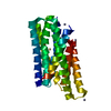
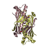
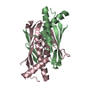
 Structure visualization
 Downloads & links
 Other downloads
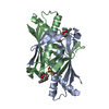


 PDBj
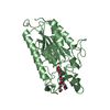
PDBj Assembly
 Mass: 18.015 Da / Num. of mol.: 285 / Source method: isolated from a natural source / Formula: H2O
 Sample preparation
 Processing