 Movie
Movie Controller

[English] 日本語
 Yorodumi
- PDB-1ldi: CRYSTAL STRUCTURE OF THE E. COLI GLYCEROL FACILITATOR (GLPF) WITH... -
+ Open data

- Basic information
| Entry | Database: PDB / ID: 1ldi | ||||||
|---|---|---|---|---|---|---|---|
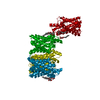
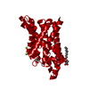
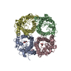
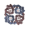
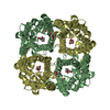
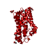
| Title | CRYSTAL STRUCTURE OF THE E. COLI GLYCEROL FACILITATOR (GLPF) WITHOUT SUBSTRATE GLYCEROL | ||||||
 Components Components | Glycerol uptake facilitator protein | ||||||
 Keywords Keywords | TRANSPORT PROTEIN / GLYCEROL-CONDUCTING MEMBRANE CHANNEL PROTEIN | ||||||
| Function / homology |  Function and homology information Function and homology informationglycerol transmembrane transporter activity / glycerol channel activity / glycerol transmembrane transport / cellular response to mercury ion / membrane => GO:0016020 / channel activity / metal ion binding / plasma membrane Similarity search - Function | ||||||
| Biological species |  | ||||||
| Method |  X-RAY DIFFRACTION / SYNCHROTRON / MOLECULAR REPLACEMENT / Resolution: 2.7 Å X-RAY DIFFRACTION / SYNCHROTRON / MOLECULAR REPLACEMENT / Resolution: 2.7 Å | ||||||
 Authors Authors | Nollert, P. / Miercke, L.J.W. / O'Connell, J. / Stroud, R.M. | ||||||
 Citation Citation | Journal: Science / Year: 2002 Title: Control of the selectivity of the aquaporin water channel family by global orientational tuning. Authors: Tajkhorshid, E. / Nollert, P. / Jensen, M.O. / Miercke, L.J. / O'Connell, J. / Stroud, R.M. / Schulten, K. #1: Journal: Science / Year: 2000Title: Structure of a Glycerol-Conducting Channel and the Basis for Its Selectivity Authors: Fu, D. / Libson, A. / Miercke, L.J. / Weitzman, C. / Nollert, P. / Krucinski, J. / Stroud, R.M. | ||||||
| History |
| ||||||
| Remark 650 | HELIX DETERMINATION METHOD: AUTHOR |
- Structure visualization
| Structure viewer | Molecule: MolmilJmol/JSmol |
|---|
- Downloads & links
-Download
| PDBx/mmCIF format | 1ldi.cif.gz | 62.4 KB | Display | PDBx/mmCIF format |
|---|---|---|---|---|
| PDB format | pdb1ldi.ent.gz | 46 KB | Display | PDB format |
| PDBx/mmJSON format | 1ldi.json.gz | Tree view | PDBx/mmJSON format | |
| Others |  Other downloads Other downloads |
-Validation report
| Arichive directory | https://data.pdbj.org/pub/pdb/validation_reports/ld/1ldiftp://data.pdbj.org/pub/pdb/validation_reports/ld/1ldi | HTTPS FTP |
|---|
-Related structure data
| Related structure data |  1ldaC  1ldfC  1fx8S C: citing same article ( S: Starting model for refinement |
|---|---|
| Similar structure data |


-Links
 PDBj
PDBj
- Assembly
| Deposited unit | 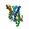
| ||||||||
|---|---|---|---|---|---|---|---|---|---|
| 1 | 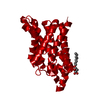
| ||||||||
| Unit cell |
| ||||||||
| Details | The biological assembly is created by the crystallographic four-fold axis: -X+1, -Y+1, Z; -Y+1,X,Z; Y,-X+1,Z |
-Components
| #1: Protein | Mass: 29799.842 Da / Num. of mol.: 1 Source method: isolated from a genetically manipulated source Source: (gene. exp.) | ||
|---|---|---|---|
| #2: Sugar |   Type: D-saccharide / Mass: 292.369 Da / Num. of mol.: 2 Type: D-saccharide / Mass: 292.369 Da / Num. of mol.: 2Source method: isolated from a genetically manipulated source Formula: C14H28O6 / Comment: detergent*YM #3: Water | ChemComp-HOH / |  Mass: 18.015 Da / Num. of mol.: 62 / Source method: isolated from a natural source / Formula: H2O Mass: 18.015 Da / Num. of mol.: 62 / Source method: isolated from a natural source / Formula: H2O |
-Experimental details
-Experiment
| Experiment | Method: X-RAY DIFFRACTION / Number of used crystals: 1 |
|---|
- Sample preparation
| Crystal | Density Matthews: 3.56 Å3/Da / Density % sol: 65.47 % | ||||||||||||||||||||||||||||||||||||||||||||||||
|---|---|---|---|---|---|---|---|---|---|---|---|---|---|---|---|---|---|---|---|---|---|---|---|---|---|---|---|---|---|---|---|---|---|---|---|---|---|---|---|---|---|---|---|---|---|---|---|---|---|
| Crystal grow | Temperature: 298 K / Method: vapor diffusion, hanging drop / pH: 9.5 Details: GLPF at 15-20 mg/ml, 100 mM Bicine, 28% (w/v) PEG 2000, 300mM MgCl2, 5 mMDTT, 15 % (v/v) xylose, 35 mM N-octyl-beta-D-glucoside, pH 9.50, VAPOR DIFFUSION, HANGING DROP, temperature 298K | ||||||||||||||||||||||||||||||||||||||||||||||||
| Crystal grow | *PLUS pH: 8.9 / Method: unknown | ||||||||||||||||||||||||||||||||||||||||||||||||
| Components of the solutions | *PLUS
|
-Data collection
| Diffraction | Mean temperature: 100 K |
|---|---|
| Diffraction source | Source: SYNCHROTRON / Site: ALS  / Beamline: 5.0.2 / Wavelength: 1.1 / Beamline: 5.0.2 / Wavelength: 1.1 |
| Detector | Type: ADSC QUANTUM / Detector: CCD / Date: Jun 7, 2000 |
| Radiation | Protocol: SINGLE WAVELENGTH / Monochromatic (M) / Laue (L): M / Scattering type: x-ray |
| Radiation wavelength | Wavelength: 1.1 Å / Relative weight: 1 |
| Reflection | Resolution: 2.7→30 Å / Num. obs: 10918 / % possible obs: 87.5 % / Observed criterion σ(I): 0 / Redundancy: 5.949 % / Rmerge(I) obs: 0.081 / Net I/σ(I): 14 |
| Reflection shell | Highest resolution: 2.7 Å / Rmerge(I) obs: 0.322 / % possible all: 83.2 |
| Reflection | *PLUS Highest resolution: 2.7 Å / Lowest resolution: 30 Å / Num. measured all: 64951 / Rmerge(I) obs: 0.081 |
| Reflection shell | *PLUS Rmerge(I) obs: 0.322 |
- Processing
| Software |
| ||||||||||||||||||||||||||||||||||||||||||||||||||||||||||||
|---|---|---|---|---|---|---|---|---|---|---|---|---|---|---|---|---|---|---|---|---|---|---|---|---|---|---|---|---|---|---|---|---|---|---|---|---|---|---|---|---|---|---|---|---|---|---|---|---|---|---|---|---|---|---|---|---|---|---|---|---|---|
| Refinement | Method to determine structure: MOLECULAR REPLACEMENT Starting model: PDB ENTRY 1FX8 Resolution: 2.7→30 Å / σ(F): 0 / Stereochemistry target values: ENGH & HUBER
| ||||||||||||||||||||||||||||||||||||||||||||||||||||||||||||
| Refinement step | Cycle: LAST / Resolution: 2.7→30 Å
| ||||||||||||||||||||||||||||||||||||||||||||||||||||||||||||
| Refine LS restraints |
| ||||||||||||||||||||||||||||||||||||||||||||||||||||||||||||
| Refinement | *PLUS Highest resolution: 2.7 Å / Lowest resolution: 30 Å / Rfactor obs: 0.229 / Rfactor Rfree: 0.261 / Rfactor Rwork: 0.229 | ||||||||||||||||||||||||||||||||||||||||||||||||||||||||||||
| Solvent computation | *PLUS | ||||||||||||||||||||||||||||||||||||||||||||||||||||||||||||
| Displacement parameters | *PLUS |
