[English] 日本語
Yorodumi
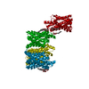
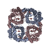
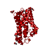
- PDB-6kxw: Crystal structure of human aquaporin AQP7 in bound to glycerol -

+
Open data


ID or keywords:

Loading...

-
Basic information

Entry
Database: PDB / ID: 6kxw
TitleCrystal structure of human aquaporin AQP7 in bound to glycerol
ComponentsAquaporin-7
KeywordsMEMBRANE PROTEIN / aquaporin / transporter / lipid metabolism
Function / homology
Function and homology information


Transport of glycerol from adipocytes to the liver by Aquaporins / glycerol channel activity / urea transmembrane transporter activity / Passive transport by Aquaporins / glycerol transmembrane transport / water transport / water channel activity / lipid droplet / cytoplasmic vesicle membrane / cell-cell junction ...Transport of glycerol from adipocytes to the liver by Aquaporins / glycerol channel activity / urea transmembrane transporter activity / Passive transport by Aquaporins / glycerol transmembrane transport / water transport / water channel activity / lipid droplet / cytoplasmic vesicle membrane / cell-cell junction / cell cortex / basolateral plasma membrane / plasma membrane / cytoplasm
Similarity search - Function
Glycerol uptake facilitator protein / Glycerol uptake facilitator protein. / Major intrinsic protein / Major intrinsic protein / Aquaporin-like / Up-down Bundle / Mainly Alpha
Similarity search - Domain/homology
Biological speciesHomo sapiens (human)
MethodX-RAY DIFFRACTION / SYNCHROTRON / MOLECULAR REPLACEMENT / Resolution: 3.7 Å
AuthorsZhang, L. / Yao, D. / Zhou, F. / Zhang, Q. / Zhou, L. / Cao, Y.
Funding support China, 4items
OrganizationGrant numberCountry
Ministry of Science and Technology (China)2017YFC1001303 China
Ministry of Science and Technology (China)2018YFC1004704 China
National Science Foundation (China)U1632132 China
National Science Foundation (China)31670849 China
CitationJournal: Sci Bull (Beijing) / Year: 2020
Title: The structural basis for glycerol permeation by human AQP7
Authors: Zhang, L. / Yao, D. / Xia, Y. / Zhang, Q. / Zhou, F. / Wang, Q. / Qin, A. / Zhao, J. / Li, D. / Li, Y. / Zhou, L. / Cao, Y.
History
DepositionSep 13, 2019Deposition site: PDBJ / Processing site: PDBJ
Revision 1.0Nov 27, 2019Provider: repository / Type: Initial release
Revision 1.1Dec 16, 2020Group: Database references / Category: citation / citation_author
Item: _citation.country / _citation.journal_abbrev ..._citation.country / _citation.journal_abbrev / _citation.journal_id_CSD / _citation.journal_id_ISSN / _citation.pdbx_database_id_DOI / _citation.title / _citation.year
Revision 1.2Sep 8, 2021Group: Database references / Category: citation / database_2
Item: _citation.journal_volume / _citation.page_first ..._citation.journal_volume / _citation.page_first / _citation.page_last / _citation.title / _database_2.pdbx_DOI / _database_2.pdbx_database_accession
Revision 1.3Nov 22, 2023Group: Data collection / Refinement description
Category: chem_comp_atom / chem_comp_bond / pdbx_initial_refinement_model

-
Structure visualization

Structure viewerMolecule:
MolmilJmol/JSmol

Downloads & links

-
Assembly

Deposited unit
A: Aquaporin-7
B: Aquaporin-7
C: Aquaporin-7
D: Aquaporin-7
hetero molecules


Theoretical massNumber of molelcules
Total (without water)149,2426
Polymers149,0584
Non-polymers1842
Water00
1


  • Idetical with deposited unit
  • defined by author&software
  • Evidence: gel filtration
TypeNameSymmetry operationNumber
identity operation1_555x,y,z1
Buried area15300 Å2
ΔGint-147 kcal/mol
Surface area35670 Å2
MethodPISA
Unit cell
Length a, b, c (Å)93.508, 106.286, 214.416
Angle α, β, γ (deg.)90.000, 90.000, 90.000
Int Tables number19
Space group name H-MP212121

-
Components

#1: Protein
Aquaporin-7 / AQP-7 / Aquaglyceroporin-7 / Aquaporin adipose / AQPap / Aquaporin-7-like


Mass: 37264.395 Da / Num. of mol.: 4
Source method: isolated from a genetically manipulated source
Source: (gene. exp.) Homo sapiens (human) / Gene: AQP7, AQP7L, AQP9 / Production host: 
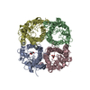
Spodoptera frugiperda (fall armyworm) / References: UniProt: O14520
#2: Chemical ChemComp-GOL / GLYCEROL / GLYCERIN / PROPANE-1,2,3-TRIOL


Mass: 92.094 Da / Num. of mol.: 2 / Source method: obtained synthetically / Formula: C3H8O3 / Feature type: SUBJECT OF INVESTIGATION
Has ligand of interestY

-
Experimental details

-
Experiment

ExperimentMethod: X-RAY DIFFRACTION / Number of used crystals: 1

-
Sample preparation

CrystalDensity Matthews: 3.57 Å3/Da / Density % sol: 65.59 %
Crystal growTemperature: 277 K / Method: evaporation
Details: 30% v/v PEG 400, 50mM CAPS, pH 10.0, 0.16% w/v 1,5-Naphthalenedisulfonic acid disodium, 0.16% w/v Naphthalene-1,3,6-trisulfonic acid trisodium, 0.16% 1,4-piperazinediethanesulfonic acid

-
Data collection

DiffractionMean temperature: 93 K / Serial crystal experiment: N
Diffraction sourceSource: SYNCHROTRON / Site: SSRF / Beamline: BL18U1 / Wavelength: 0.97775 Å
DetectorType: DECTRIS PILATUS3 6M / Detector: PIXEL / Date: Mar 16, 2019
RadiationProtocol: SINGLE WAVELENGTH / Monochromatic (M) / Laue (L): M / Scattering type: x-ray
Radiation wavelengthWavelength: 0.97775 Å / Relative weight: 1
ReflectionResolution: 3.7→47.614 Å / Num. obs: 16925 / % possible obs: 99.9 % / Redundancy: 20 % / CC1/2: 0.973 / Net I/σ(I): 9.05
Reflection shellResolution: 3.7→3.8333 Å / Num. unique obs: 311 / CC1/2: 0.608

-
Processing

Software
NameVersionClassification
PHENIX1.13_2998refinement
PDB_EXTRACT3.25data extraction
HKL-2000data reduction
HKL-2000data scaling
PHENIXphasing
RefinementMethod to determine structure: MOLECULAR REPLACEMENT
Starting model: 6F7H
Resolution: 3.7→47.614 Å / SU ML: 0.6 / Cross valid method: THROUGHOUT / σ(F): 1.35 / Phase error: 34.36
RfactorNum. reflection% reflection
Rfree0.3221 881 5.22 %
Rwork0.2679 --
obs0.2707 16881 71.86 %
Solvent computationShrinkage radii: 0.9 Å / VDW probe radii: 1.11 Å
Displacement parametersBiso max: 268.64 Å2 / Biso mean: 109.2368 Å2 / Biso min: 7.27 Å2
Refinement stepCycle: final / Resolution: 3.7→47.614 Å
ProteinNucleic acidLigandSolventTotal
Num. atoms7574 0 12 0 7586
Biso mean--55.2 --
Num. residues----999
LS refinement shell

Refine-ID: X-RAY DIFFRACTION / Rfactor Rfree error: 0

Resolution (Å)Rfactor RfreeNum. reflection RfreeRfactor RworkNum. reflection Rwork% reflection obs (%)
3.7004-3.93220.333637626
3.9322-4.23560.366482144940
4.2356-4.66150.32121350.2547270473
4.6615-5.33530.31882020.2461366699
5.3353-6.71890.38742270.3063712100
6.7189-47.610.2711980.2655384399
Refinement TLS params.Method: refined / Origin x: -13.1329 Å / Origin y: -44.6983 Å / Origin z: 21.6915 Å
111213212223313233
T0.2585 Å20.0071 Å2-0.0292 Å2-0.6448 Å20.5975 Å2--0.5005 Å2
L0.971 °2-0.0555 °2-0.1248 °2-0.9937 °20.0053 °2--1.0354 °2
S0.2753 Å °-1.72 Å °-1.1495 Å °0.1902 Å °-0.269 Å °0.0275 Å °0.0967 Å °0.1392 Å °0.0035 Å °
Refinement TLS group
IDRefine-IDRefine TLS-IDSelection detailsAuth asym-IDAuth seq-ID
1X-RAY DIFFRACTION1allA27 - 401
2X-RAY DIFFRACTION1allB30 - 278
3X-RAY DIFFRACTION1allC30 - 400
4X-RAY DIFFRACTION1allD30 - 278

+
About Yorodumi

-
News

-
Feb 9, 2022. New format data for meta-information of EMDB entries

New format data for meta-information of EMDB entries

  • Version 3 of the EMDB header file is now the official format.
  • The previous official version 1.9 will be removed from the archive.

Related info.:EMDB header

External links:wwPDB to switch to version 3 of the EMDB data model

-
Aug 12, 2020. Covid-19 info

Covid-19 info

URL: https://pdbjlvh1.pdbj.org/emnavi/covid19.php

New page: Covid-19 featured information page in EM Navigator.

Related info.:Covid-19 info / Mar 5, 2020. Novel coronavirus structure data

+
Mar 5, 2020. Novel coronavirus structure data

Novel coronavirus structure data

Related info.:Yorodumi Speices / Aug 12, 2020. Covid-19 info

External links:COVID-19 featured content - PDBj / Molecule of the Month (242):Coronavirus Proteases

+
Jan 31, 2019. EMDB accession codes are about to change! (news from PDBe EMDB page)

EMDB accession codes are about to change! (news from PDBe EMDB page)

  • The allocation of 4 digits for EMDB accession codes will soon come to an end. Whilst these codes will remain in use, new EMDB accession codes will include an additional digit and will expand incrementally as the available range of codes is exhausted. The current 4-digit format prefixed with “EMD-” (i.e. EMD-XXXX) will advance to a 5-digit format (i.e. EMD-XXXXX), and so on. It is currently estimated that the 4-digit codes will be depleted around Spring 2019, at which point the 5-digit format will come into force.
  • The EM Navigator/Yorodumi systems omit the EMD- prefix.

Related info.:Q: What is EMD? / ID/Accession-code notation in Yorodumi/EM Navigator

External links:EMDB Accession Codes are Changing Soon! / Contact to PDBj

+
Jul 12, 2017. Major update of PDB

Major update of PDB

  • wwPDB released updated PDB data conforming to the new PDBx/mmCIF dictionary.
  • This is a major update changing the version number from 4 to 5, and with Remediation, in which all the entries are updated.
  • In this update, many items about electron microscopy experimental information are reorganized (e.g. em_software).
  • Now, EM Navigator and Yorodumi are based on the updated data.

External links:wwPDB Remediation / Enriched Model Files Conforming to OneDep Data Standards Now Available in the PDB FTP Archive

-
Yorodumi

Thousand views of thousand structures

  • Yorodumi is a browser for structure data from EMDB, PDB, SASBDB, etc.
  • This page is also the successor to EM Navigator detail page, and also detail information page/front-end page for Omokage search.
  • The word "yorodu" (or yorozu) is an old Japanese word meaning "ten thousand". "mi" (miru) is to see.

Related info.:EMDB / PDB / SASBDB / Comparison of 3 databanks / Yorodumi Search / Aug 31, 2016. New EM Navigator & Yorodumi / Yorodumi Papers / Jmol/JSmol / Function and homology information / Changes in new EM Navigator and Yorodumi

Read more