 Movie
Movie Controller

[English] 日本語
 Yorodumi
- PDB-4xld: Crystal structure of the human PPARg-LBD/rosiglitazone complex ob... -
+ Open data

- Basic information
| Entry | Database: PDB / ID: 4xld | ||||||
|---|---|---|---|---|---|---|---|
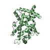
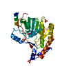
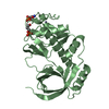
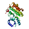
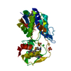
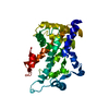
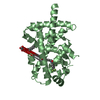
| Title | Crystal structure of the human PPARg-LBD/rosiglitazone complex obtained by dry co-crystallization and in situ diffraction | ||||||
 Components Components | Peroxisome proliferator-activated receptor gamma | ||||||
 Keywords Keywords | GENE REGULATION / nuclear receptor ligand screening | ||||||
| Function / homology |  Function and homology information Function and homology informationprostaglandin receptor activity / negative regulation of receptor signaling pathway via STAT / MECP2 regulates transcription factors / beige fat cell differentiation / negative regulation of vascular endothelial cell proliferation / negative regulation of extracellular matrix assembly / negative regulation of connective tissue replacement involved in inflammatory response wound healing / positive regulation of cholesterol transport / negative regulation of cellular response to transforming growth factor beta stimulus / arachidonate binding ...prostaglandin receptor activity / negative regulation of receptor signaling pathway via STAT / MECP2 regulates transcription factors / beige fat cell differentiation / negative regulation of vascular endothelial cell proliferation / negative regulation of extracellular matrix assembly / negative regulation of connective tissue replacement involved in inflammatory response wound healing / positive regulation of cholesterol transport / negative regulation of cellular response to transforming growth factor beta stimulus / arachidonate binding / positive regulation of adiponectin secretion / DNA binding domain binding / positive regulation of vascular associated smooth muscle cell apoptotic process / negative regulation of cardiac muscle hypertrophy in response to stress / positive regulation of fatty acid metabolic process / positive regulation of lipid metabolic process / STAT family protein binding / WW domain binding / negative regulation of type II interferon-mediated signaling pathway / LBD domain binding / negative regulation of cholesterol storage / response to lipid / positive regulation of lipoprotein transport / negative regulation of SMAD protein signal transduction / lipid homeostasis / E-box binding / R-SMAD binding / negative regulation of blood vessel endothelial cell migration / white fat cell differentiation / negative regulation of vascular associated smooth muscle cell proliferation / alpha-actinin binding / negative regulation of macrophage derived foam cell differentiation / negative regulation of lipid storage / positive regulation of cholesterol efflux / monocyte differentiation / negative regulation of BMP signaling pathway / cell fate commitment / cellular response to low-density lipoprotein particle stimulus / long-chain fatty acid transport / BMP signaling pathway / negative regulation of mitochondrial fission / negative regulation of osteoblast differentiation / nuclear retinoid X receptor binding / positive regulation of fat cell differentiation / fat cell differentiation / Transcriptional regulation of brown and beige adipocyte differentiation by EBF2 / retinoic acid receptor signaling pathway / intracellular receptor signaling pathway / negative regulation of MAPK cascade / cell maturation / peptide binding / peroxisome proliferator activated receptor signaling pathway / epithelial cell differentiation / hormone-mediated signaling pathway / regulation of cellular response to insulin stimulus / response to nutrient / positive regulation of adipose tissue development / negative regulation of miRNA transcription / negative regulation of angiogenesis / brown fat cell differentiation / placenta development / Regulation of PTEN gene transcription / transcription coregulator binding / SUMOylation of intracellular receptors / positive regulation of apoptotic signaling pathway / negative regulation of smooth muscle cell proliferation / negative regulation of transforming growth factor beta receptor signaling pathway / PPARA activates gene expression / fatty acid metabolic process / Nuclear Receptor transcription pathway / Transcriptional regulation of white adipocyte differentiation / regulation of circadian rhythm / positive regulation of miRNA transcription / DNA-binding transcription repressor activity, RNA polymerase II-specific / mRNA transcription by RNA polymerase II / nuclear receptor activity / negative regulation of inflammatory response / regulation of blood pressure / RNA polymerase II transcription regulator complex / cellular response to insulin stimulus / rhythmic process / glucose homeostasis / MLL4 and MLL3 complexes regulate expression of PPARG target genes in adipogenesis and hepatic steatosis / double-stranded DNA binding / DNA-binding transcription activator activity, RNA polymerase II-specific / cellular response to hypoxia / sequence-specific DNA binding / DNA-binding transcription factor binding / nucleic acid binding / DNA-binding transcription factor activity, RNA polymerase II-specific / cell differentiation / signaling receptor complex / transcription cis-regulatory region binding / RNA polymerase II cis-regulatory region sequence-specific DNA binding / DNA-binding transcription factor activity / negative regulation of gene expression / innate immune response / negative regulation of DNA-templated transcription / chromatin binding / positive regulation of gene expression Similarity search - Function | ||||||
| Biological species |  Homo sapiens (human) Homo sapiens (human) | ||||||
| Method |  X-RAY DIFFRACTION / SYNCHROTRON / MOLECULAR REPLACEMENT / Resolution: 2.45 Å X-RAY DIFFRACTION / SYNCHROTRON / MOLECULAR REPLACEMENT / Resolution: 2.45 Å | ||||||
 Authors Authors | Delfosse, V. / Guichou, J.-F. | ||||||
| Funding support |  France, 1items France, 1items
| ||||||
 Citation Citation | Journal: Acta Crystallogr.,Sect.D / Year: 2015 Title: Combining `dry' co-crystallization and in situ diffraction to facilitate ligand screening by X-ray crystallography. Authors: Gelin, M. / Delfosse, V. / Allemand, F. / Hoh, F. / Sallaz-Damaz, Y. / Pirocchi, M. / Bourguet, W. / Ferrer, J.L. / Labesse, G. / Guichou, J.F. | ||||||
| History |
|
- Structure visualization
| Structure viewer | Molecule: MolmilJmol/JSmol |
|---|
- Downloads & links
-Download
| PDBx/mmCIF format | 4xld.cif.gz | 67 KB | Display | PDBx/mmCIF format |
|---|---|---|---|---|
| PDB format | pdb4xld.ent.gz | 47.8 KB | Display | PDB format |
| PDBx/mmJSON format | 4xld.json.gz | Tree view | PDBx/mmJSON format | |
| Others |  Other downloads Other downloads |
-Validation report
| Arichive directory | https://data.pdbj.org/pub/pdb/validation_reports/xl/4xldftp://data.pdbj.org/pub/pdb/validation_reports/xl/4xld | HTTPS FTP |
|---|
-Related structure data
| Related structure data |  3rdcC  4xn6C  4xncC  4xneC  4xoyC  4xozC  4xp0C  4xp2C  4xp3C  4xrjC  4xrlC  4zscC 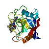 4zsdC 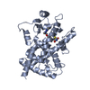 2prgS S: Starting model for refinement C: citing same article ( |
|---|---|
| Similar structure data |


-Links
 PDBj
PDBj- Assembly
| Deposited unit | 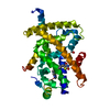
| ||||||||
|---|---|---|---|---|---|---|---|---|---|
| 1 |
| ||||||||
| Unit cell |
|
-Components
| #1: Protein | Mass: 33752.066 Da / Num. of mol.: 1 / Fragment: UNP residues 203-477 Source method: isolated from a genetically manipulated source Source: (gene. exp.) Homo sapiens (human) / Gene: PPARG, NR1C3 / Plasmid: pET15b / Production host:  |
|---|---|
| #2: Chemical | ChemComp-FMT /   Mass: 46.025 Da / Num. of mol.: 1 / Source method: obtained synthetically / Formula: CH2O2 Mass: 46.025 Da / Num. of mol.: 1 / Source method: obtained synthetically / Formula: CH2O2 |
| #3: Chemical | ChemComp-BRL /   Mass: 357.427 Da / Num. of mol.: 1 / Source method: obtained synthetically / Formula: C18H19N3O3S Mass: 357.427 Da / Num. of mol.: 1 / Source method: obtained synthetically / Formula: C18H19N3O3S |
| #4: Water | ChemComp-HOH /  Mass: 18.015 Da / Num. of mol.: 25 / Source method: isolated from a natural source / Formula: H2O Mass: 18.015 Da / Num. of mol.: 25 / Source method: isolated from a natural source / Formula: H2O |
-Experimental details
-Experiment
| Experiment | Method: X-RAY DIFFRACTION |
|---|
- Sample preparation
| Crystal | Density Matthews: 2.54 Å3/Da / Density % sol: 51 % |
|---|---|
| Crystal grow | Temperature: 291 K / Method: vapor diffusion, sitting drop / pH: 8 / Details: 3-4 M Sodium Formate / PH range: 7-7.5 |
-Data collection
| Diffraction | Mean temperature: 291 K Ambient temp details: in situ (in plate), at room temperature |
|---|---|
| Diffraction source | Source: SYNCHROTRON / Site: ESRF / Beamline: BM30A / Wavelength: 0.97922 Å |
| Detector | Type: ADSC QUANTUM 315r / Detector: CCD / Date: Jul 21, 2014 |
| Radiation | Protocol: SINGLE WAVELENGTH / Monochromatic (M) / Laue (L): M / Scattering type: x-ray |
| Radiation wavelength | Wavelength: 0.97922 Å / Relative weight: 1 |
| Reflection | Resolution: 2.45→47.37 Å / Num. obs: 12488 / % possible obs: 90 % / Redundancy: 4.4 % / Biso Wilson estimate: 41.1 Å2 / Rsym value: 0.154 / Net I/σ(I): 7.43 |
| Reflection shell | Resolution: 2.45→2.59 Å / Redundancy: 3.7 % / Rmerge(I) obs: 0.477 / Mean I/σ(I) obs: 4.68 / % possible all: 77.8 |
- Processing
| Software |
| ||||||||||||||||||||||||||||||||||||||||||||||||||||||||
|---|---|---|---|---|---|---|---|---|---|---|---|---|---|---|---|---|---|---|---|---|---|---|---|---|---|---|---|---|---|---|---|---|---|---|---|---|---|---|---|---|---|---|---|---|---|---|---|---|---|---|---|---|---|---|---|---|---|
| Refinement | Method to determine structure: MOLECULAR REPLACEMENT Starting model: 2prg Resolution: 2.45→47.369 Å / SU ML: 0.25 / Cross valid method: FREE R-VALUE / σ(F): 1.36 / Phase error: 22.15 / Stereochemistry target values: ML
| ||||||||||||||||||||||||||||||||||||||||||||||||||||||||
| Solvent computation | Shrinkage radii: 0.9 Å / VDW probe radii: 1.11 Å / Solvent model: FLAT BULK SOLVENT MODEL | ||||||||||||||||||||||||||||||||||||||||||||||||||||||||
| Displacement parameters | Biso mean: 44.7 Å2 | ||||||||||||||||||||||||||||||||||||||||||||||||||||||||
| Refinement step | Cycle: LAST / Resolution: 2.45→47.369 Å
| ||||||||||||||||||||||||||||||||||||||||||||||||||||||||
| Refine LS restraints |
| ||||||||||||||||||||||||||||||||||||||||||||||||||||||||
| LS refinement shell |
|
