 Movie
Movie Controller

[English] 日本語
 Yorodumi
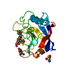
- PDB-4zsc: Human Cyclophilin D Complexed with an Inhibitor at room temperature -
+ Open data

- Basic information
| Entry | Database: PDB / ID: 4zsc | ||||||
|---|---|---|---|---|---|---|---|
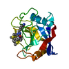
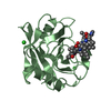
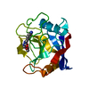
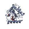
| Title | Human Cyclophilin D Complexed with an Inhibitor at room temperature | ||||||
 Components Components | Peptidyl-prolyl cis-trans isomerase F, mitochondrial | ||||||
 Keywords Keywords | ISOMERASE / isomerase inhibitor complex | ||||||
| Function / homology |  Function and homology information Function and homology information: / mitochondrial outer membrane permeabilization involved in programmed cell death / regulation of mitochondrial membrane permeability involved in programmed necrotic cell death / skeletal muscle fiber differentiation / mitochondrial permeability transition pore complex / cellular response to arsenic-containing substance / mitochondrial depolarization / negative regulation of oxidative phosphorylation / regulation of mitochondrial membrane permeability / cyclosporin A binding ...: / mitochondrial outer membrane permeabilization involved in programmed cell death / regulation of mitochondrial membrane permeability involved in programmed necrotic cell death / skeletal muscle fiber differentiation / mitochondrial permeability transition pore complex / cellular response to arsenic-containing substance / mitochondrial depolarization / negative regulation of oxidative phosphorylation / regulation of mitochondrial membrane permeability / cyclosporin A binding / negative regulation of release of cytochrome c from mitochondria / necroptotic process / negative regulation of intrinsic apoptotic signaling pathway / apoptotic mitochondrial changes / cellular response to calcium ion / response to ischemia / peptidylprolyl isomerase / peptidyl-prolyl cis-trans isomerase activity / cellular response to hydrogen peroxide / protein folding / mitochondrial matrix / negative regulation of apoptotic process / mitochondrion / membrane / cytoplasm Similarity search - Function | ||||||
| Biological species |  Homo sapiens (human) Homo sapiens (human) | ||||||
| Method |  X-RAY DIFFRACTION / SYNCHROTRON / Resolution: 1.5 Å X-RAY DIFFRACTION / SYNCHROTRON / Resolution: 1.5 Å | ||||||
 Authors Authors | Gelin, M. / Delfosse, V. / Allemand, F. / Hoh, F. / Sallaz-Damaz, Y. / Pirocchi, M. / Bourguet, W. / Ferrer, J.-L. / Labesse, G. / Guichou, J.-F. | ||||||
 Citation Citation | Journal: Acta Crystallogr.,Sect.D / Year: 2015 Title: Combining `dry' co-crystallization and in situ diffraction to facilitate ligand screening by X-ray crystallography. Authors: Gelin, M. / Delfosse, V. / Allemand, F. / Hoh, F. / Sallaz-Damaz, Y. / Pirocchi, M. / Bourguet, W. / Ferrer, J.L. / Labesse, G. / Guichou, J.F. | ||||||
| History |
|
- Structure visualization
| Structure viewer | Molecule: MolmilJmol/JSmol |
|---|
- Downloads & links
-Download
| PDBx/mmCIF format | 4zsc.cif.gz | 77.6 KB | Display | PDBx/mmCIF format |
|---|---|---|---|---|
| PDB format | pdb4zsc.ent.gz | 57.2 KB | Display | PDB format |
| PDBx/mmJSON format | 4zsc.json.gz | Tree view | PDBx/mmJSON format | |
| Others |  Other downloads Other downloads |
-Validation report
| Arichive directory | https://data.pdbj.org/pub/pdb/validation_reports/zs/4zscftp://data.pdbj.org/pub/pdb/validation_reports/zs/4zsc | HTTPS FTP |
|---|
-Related structure data
| Related structure data |  3rdcC  4xldC  4xn6C  4xncC  4xneC  4xoyC  4xozC  4xp0C  4xp2C  4xp3C  4xrjC  4xrlC  4zsdC C: citing same article ( |
|---|---|
| Similar structure data |


-Links
 PDBj
PDBj

- Assembly
| Deposited unit | 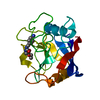
| ||||||||
|---|---|---|---|---|---|---|---|---|---|
| 1 |
| ||||||||
| Unit cell |
|
-Components
| #1: Protein | Mass: 17652.125 Da / Num. of mol.: 1 / Fragment: UNP residues 44-207 / Mutation: K133I Source method: isolated from a genetically manipulated source Source: (gene. exp.) Homo sapiens (human) / Gene: PPIF, CYP3 / Production host:  |
|---|---|
| #2: Chemical | ChemComp-EA4 /   Mass: 251.282 Da / Num. of mol.: 1 / Source method: obtained synthetically / Formula: C12H17N3O3 Mass: 251.282 Da / Num. of mol.: 1 / Source method: obtained synthetically / Formula: C12H17N3O3 |
| #3: Water | ChemComp-HOH /  Mass: 18.015 Da / Num. of mol.: 91 / Source method: isolated from a natural source / Formula: H2O Mass: 18.015 Da / Num. of mol.: 91 / Source method: isolated from a natural source / Formula: H2O |
-Experimental details
-Experiment
| Experiment | Method: X-RAY DIFFRACTION / Number of used crystals: 1 |
|---|
- Sample preparation
| Crystal | Density Matthews: 2.1 Å3/Da / Density % sol: 41.36 % |
|---|---|
| Crystal grow | Temperature: 291 K / Method: vapor diffusion, sitting drop / pH: 7.3 / Details: 30% PEG4000 |
-Data collection
| Diffraction | Mean temperature: 298 K |
|---|---|
| Diffraction source | Source: SYNCHROTRON / Site: ESRF  / Beamline: BM30A / Wavelength: 0.9797 Å / Beamline: BM30A / Wavelength: 0.9797 Å |
| Detector | Type: ADSC QUANTUM 315r / Detector: CCD / Date: May 7, 2015 |
| Radiation | Protocol: SINGLE WAVELENGTH / Monochromatic (M) / Laue (L): M / Scattering type: x-ray |
| Radiation wavelength | Wavelength: 0.9797 Å / Relative weight: 1 |
| Reflection | Limit h max: 25 / Limit h min: 1 / Limit k max: 18 / Limit k min: 0 / Limit l max: 38 / Limit l min: 0 / Number: 53929 / D res high: 2.233 Å / D res low: 29 Å / Num. obs: 53901 |
| Reflection | Resolution: 1.5→48.4 Å / Num. obs: 16982 / % possible obs: 73.1 % / Redundancy: 2.8 % / Net I/σ(I): 12.1 |
| Reflection scale | Group code: 1 |
- Processing
| Software |
| ||||||||||||||||||||||||||||||||||||||||||||||||||||||||||||||||||||||||||||||||||||||||||
|---|---|---|---|---|---|---|---|---|---|---|---|---|---|---|---|---|---|---|---|---|---|---|---|---|---|---|---|---|---|---|---|---|---|---|---|---|---|---|---|---|---|---|---|---|---|---|---|---|---|---|---|---|---|---|---|---|---|---|---|---|---|---|---|---|---|---|---|---|---|---|---|---|---|---|---|---|---|---|---|---|---|---|---|---|---|---|---|---|---|---|---|
| Refinement | Resolution: 1.5→48.43 Å / Cor.coef. Fo:Fc: 0.975 / Cor.coef. Fo:Fc free: 0.959 / SU B: 2.223 / SU ML: 0.037 / Cross valid method: THROUGHOUT / σ(F): 0 / ESU R: 0.123 / ESU R Free: 0.079 / Stereochemistry target values: MAXIMUM LIKELIHOOD Details: HYDROGENS HAVE BEEN ADDED IN THE RIDING POSITIONS U VALUES : REFINED INDIVIDUALLY
| ||||||||||||||||||||||||||||||||||||||||||||||||||||||||||||||||||||||||||||||||||||||||||
| Solvent computation | Ion probe radii: 0.8 Å / Shrinkage radii: 0.8 Å / VDW probe radii: 1.2 Å / Solvent model: MASK | ||||||||||||||||||||||||||||||||||||||||||||||||||||||||||||||||||||||||||||||||||||||||||
| Displacement parameters | Biso max: 35.33 Å2 / Biso mean: 9.817 Å2 / Biso min: 4.47 Å2
| ||||||||||||||||||||||||||||||||||||||||||||||||||||||||||||||||||||||||||||||||||||||||||
| Refinement step | Cycle: final / Resolution: 1.5→48.43 Å
| ||||||||||||||||||||||||||||||||||||||||||||||||||||||||||||||||||||||||||||||||||||||||||
| Refine LS restraints |
| ||||||||||||||||||||||||||||||||||||||||||||||||||||||||||||||||||||||||||||||||||||||||||
| LS refinement shell | Resolution: 1.5→1.539 Å / Total num. of bins used: 20
|
