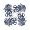
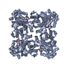
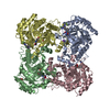
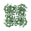
- PDB-4x3z: Inosine 5'-monophosphate dehydrogenase from Vibrio cholerae, dele... -
+
Open data
ID or keywords:
Loading...
-
Basic information
Entry
Database: PDB / ID: 4x3z
Title
Inosine 5'-monophosphate dehydrogenase from Vibrio cholerae, deletion mutant, in complex with XMP and NAD
Components
Inosine-5'-monophosphate dehydrogenase
Keywords
OXIDOREDUCTASE / INOSINE 5'-MONOPHOSPHATE DEHYDROGENASE / IMPDH / XMP / xanthosine monophosphate / structural genomics / Center for Structural Genomics of Infectious Diseases / CSGID
Function / homology
Function and homology information
IMP dehydrogenase / IMP dehydrogenase activity / GMP biosynthetic process / GTP biosynthetic process / nucleotide binding / metal ion binding Similarity search - Function
IMP dehydrogenase / GMP reductase, conserved site / IMP dehydrogenase / GMP reductase signature. / Inosine-5'-monophosphate dehydrogenase / IMP dehydrogenase/GMP reductase / IMP dehydrogenase / GMP reductase domain / IMP dehydrogenase / GMP reductase domain / Domain in cystathionine beta-synthase and other proteins. / CBS domain superfamily / CBS domain / CBS domain ...IMP dehydrogenase / GMP reductase, conserved site / IMP dehydrogenase / GMP reductase signature. / Inosine-5'-monophosphate dehydrogenase / IMP dehydrogenase/GMP reductase / IMP dehydrogenase / GMP reductase domain / IMP dehydrogenase / GMP reductase domain / Domain in cystathionine beta-synthase and other proteins. / CBS domain superfamily / CBS domain / CBS domain / CBS domain profile. / Aldolase class I / Aldolase-type TIM barrel / TIM Barrel / Alpha-Beta Barrel / Alpha Beta Similarity search - Domain/homology
Resolution: 1.62→44.2 Å / Cor.coef. Fo:Fc: 0.972 / Cor.coef. Fo:Fc free: 0.961 / SU B: 2.796 / SU ML: 0.048 / Cross valid method: THROUGHOUT / ESU R: 0.076 / ESU R Free: 0.078 / Stereochemistry target values: MAXIMUM LIKELIHOOD / Details: HYDROGENS HAVE BEEN ADDED IN THE RIDING POSITIONS
Rfactor
Num. reflection
% reflection
Selection details
Rfree
0.18157
4552
4.9 %
RANDOM
Rwork
0.152
-
-
-
obs
0.15345
87862
99.92 %
-
Solvent computation
Ion probe radii: 0.8 Å / Shrinkage radii: 0.8 Å / VDW probe radii: 1.2 Å / Solvent model: MASK
 Movie
Movie Controller

 Yorodumi
 Open data
 Basic information
 Components
 Keywords
 Function and homology information
 Vibrio cholerae O1 biovar El Tor str. N16961 (bacteria)
 X-RAY DIFFRACTION /
  Authors
 Citation
 Structure visualization
 Downloads & links
 Other downloads
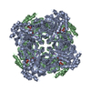


 PDBj
PDBj
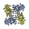
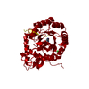
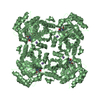
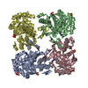
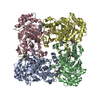
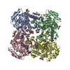
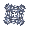
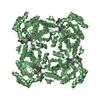
 Assembly
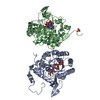
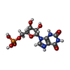


 Mass: 365.213 Da / Num. of mol.: 2 / Source method: obtained synthetically / Formula: C10H14N4O9P
 Mass: 663.425 Da / Num. of mol.: 2 / Source method: obtained synthetically / Formula: C21H27N7O14P2 / Comment: NAD*YM
 Mass: 39.098 Da / Num. of mol.: 2 / Source method: obtained synthetically / Formula: K
 Mass: 92.094 Da / Num. of mol.: 1 / Source method: obtained synthetically / Formula: C3H8O3
 Mass: 94.971 Da / Num. of mol.: 1 / Source method: obtained synthetically / Formula: PO4
 Sample preparation
 / Beamline: 19-ID / Wavelength: 0.9792 Å
 Processing