- PDB-2cu0: Crystal structure of inosine-5'-monophosphate dehydrogenase from ... -
+
Open data
ID or keywords:
Loading...
-
Basic information
Entry
Database: PDB / ID: 2cu0
Title
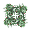
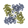
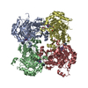
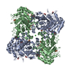
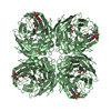
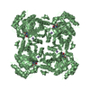
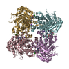
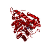
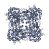
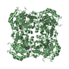
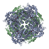
Crystal structure of inosine-5'-monophosphate dehydrogenase from Pyrococcus horikoshii OT3
Components
Inosine-5'-monophosphate dehydrogenase
Keywords
OXIDOREDUCTASE / structural genomics / Pyrococcus horikoshii OT3 / inosine-5'-monophosphate dehydrogenase / RIKEN Structural Genomics/Proteomics Initiative / RSGI / NPPSFA / National Project on Protein Structural and Functional Analyses
Function / homology
Function and homology information
IMP dehydrogenase / IMP dehydrogenase activity / GMP biosynthetic process / GTP biosynthetic process / nucleotide binding / metal ion binding Similarity search - Function
IMP dehydrogenase / GMP reductase, conserved site / IMP dehydrogenase / GMP reductase signature. / Inosine-5'-monophosphate dehydrogenase / IMP dehydrogenase/GMP reductase / IMP dehydrogenase / GMP reductase domain / IMP dehydrogenase / GMP reductase domain / Domain in cystathionine beta-synthase and other proteins. / CBS domain superfamily / CBS domain / CBS domain ...IMP dehydrogenase / GMP reductase, conserved site / IMP dehydrogenase / GMP reductase signature. / Inosine-5'-monophosphate dehydrogenase / IMP dehydrogenase/GMP reductase / IMP dehydrogenase / GMP reductase domain / IMP dehydrogenase / GMP reductase domain / Domain in cystathionine beta-synthase and other proteins. / CBS domain superfamily / CBS domain / CBS domain / CBS domain profile. / Aldolase class I / Aldolase-type TIM barrel / TIM Barrel / Alpha-Beta Barrel / Alpha Beta Similarity search - Domain/homology
The biological assembly is tetramer generated from the chain A in the asymmetric unit by the operations: x,y,z : -x,-y,z : -y,x,z : y,-x ,z / The biological assembly is tetramer generated from the chain B in the asymmetric unit by the operations: x,y,z : -x,-y,z : -y,x,z : y,-x ,z
In the structure databanks used in Yorodumi, some data are registered as the other names, "COVID-19 virus" and "2019-nCoV". Here are the details of the virus and the list of structure data.
Jan 31, 2019. EMDB accession codes are about to change! (news from PDBe EMDB page)
EMDB accession codes are about to change! (news from PDBe EMDB page)
The allocation of 4 digits for EMDB accession codes will soon come to an end. Whilst these codes will remain in use, new EMDB accession codes will include an additional digit and will expand incrementally as the available range of codes is exhausted. The current 4-digit format prefixed with “EMD-” (i.e. EMD-XXXX) will advance to a 5-digit format (i.e. EMD-XXXXX), and so on. It is currently estimated that the 4-digit codes will be depleted around Spring 2019, at which point the 5-digit format will come into force.
The EM Navigator/Yorodumi systems omit the EMD- prefix.
Related info.:Q: What is EMD? / ID/Accession-code notation in Yorodumi/EM Navigator
Yorodumi is a browser for structure data from EMDB, PDB, SASBDB, etc.
This page is also the successor to EM Navigator detail page, and also detail information page/front-end page for Omokage search.
The word "yorodu" (or yorozu) is an old Japanese word meaning "ten thousand". "mi" (miru) is to see.
Related info.:EMDB / PDB / SASBDB / Comparison of 3 databanks / Yorodumi Search / Aug 31, 2016. New EM Navigator & Yorodumi / Yorodumi Papers / Jmol/JSmol / Function and homology information / Changes in new EM Navigator and Yorodumi
 Movie
Movie Controller

 Yorodumi
 Open data
 Basic information
 Components
 Keywords
 Function and homology information
 Pyrococcus horikoshii (archaea)
 X-RAY DIFFRACTION /
  Authors
 Citation
 Structure visualization
 Downloads & links
 Other downloads
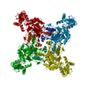


 PDBj
PDBj
 Assembly
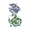

 Mass: 365.213 Da / Num. of mol.: 2 / Source method: obtained synthetically / Formula: C10H14N4O9P
 Mass: 18.015 Da / Num. of mol.: 374 / Source method: isolated from a natural source / Formula: H2O
 Sample preparation
 / Beamline: BL26B1 / Wavelength: 1 Å
 Processing