 Movie
Movie Controller


+ Open data

- Basic information
| Entry | Database: PDB / ID: 4wau | ||||||
|---|---|---|---|---|---|---|---|
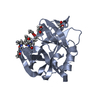
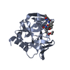
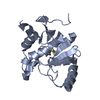
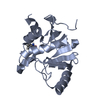
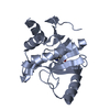
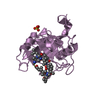
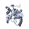
| Title | Crystal structure of CENP-M solved by native-SAD phasing | ||||||
 Components Components | Centromere protein M | ||||||
 Keywords Keywords | CELL CYCLE / native-SAD / S-SAD / sulfur-SAD / sulfur SAD / Mitosis / Kinetochore / CCAN / G-protein | ||||||
| Function / homology |  Function and homology information Function and homology informationAmplification of signal from unattached kinetochores via a MAD2 inhibitory signal / Mitotic Prometaphase / Deposition of new CENPA-containing nucleosomes at the centromere / EML4 and NUDC in mitotic spindle formation / Resolution of Sister Chromatid Cohesion / chromosome segregation / RHO GTPases Activate Formins / kinetochore / Separation of Sister Chromatids / nucleoplasm ...Amplification of signal from unattached kinetochores via a MAD2 inhibitory signal / Mitotic Prometaphase / Deposition of new CENPA-containing nucleosomes at the centromere / EML4 and NUDC in mitotic spindle formation / Resolution of Sister Chromatid Cohesion / chromosome segregation / RHO GTPases Activate Formins / kinetochore / Separation of Sister Chromatids / nucleoplasm / nucleus / cytosol Similarity search - Function | ||||||
| Biological species |  Homo sapiens (human) Homo sapiens (human) | ||||||
| Method |  X-RAY DIFFRACTION / SYNCHROTRON / SAD / Resolution: 2.2 Å X-RAY DIFFRACTION / SYNCHROTRON / SAD / Resolution: 2.2 Å | ||||||
 Authors Authors | Weinert, T. / Basilico, F. / Cecatiello, V. / Pasqualato, S. / Wang, M. | ||||||
 Citation Citation | Journal: Nat.Methods / Year: 2015 Title: Fast native-SAD phasing for routine macromolecular structure determination. Authors: Weinert, T. / Olieric, V. / Waltersperger, S. / Panepucci, E. / Chen, L. / Zhang, H. / Zhou, D. / Rose, J. / Ebihara, A. / Kuramitsu, S. / Li, D. / Howe, N. / Schnapp, G. / Pautsch, A. / ...Authors: Weinert, T. / Olieric, V. / Waltersperger, S. / Panepucci, E. / Chen, L. / Zhang, H. / Zhou, D. / Rose, J. / Ebihara, A. / Kuramitsu, S. / Li, D. / Howe, N. / Schnapp, G. / Pautsch, A. / Bargsten, K. / Prota, A.E. / Surana, P. / Kottur, J. / Nair, D.T. / Basilico, F. / Cecatiello, V. / Pasqualato, S. / Boland, A. / Weichenrieder, O. / Wang, B.C. / Steinmetz, M.O. / Caffrey, M. / Wang, M. | ||||||
| History |
|
- Structure visualization
| Structure viewer | Molecule: MolmilJmol/JSmol |
|---|
- Downloads & links
-Download
| PDBx/mmCIF format | 4wau.cif.gz | 79.1 KB | Display | PDBx/mmCIF format |
|---|---|---|---|---|
| PDB format | pdb4wau.ent.gz | 58.7 KB | Display | PDB format |
| PDBx/mmJSON format | 4wau.json.gz | Tree view | PDBx/mmJSON format | |
| Others |  Other downloads Other downloads |
-Validation report
| Arichive directory | https://data.pdbj.org/pub/pdb/validation_reports/wa/4wauftp://data.pdbj.org/pub/pdb/validation_reports/wa/4wau | HTTPS FTP |
|---|
-Related structure data
| Related structure data | 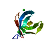 4pgoC 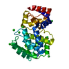 4piiC  4r8tC 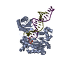 4r8uC 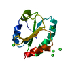 4tn8C 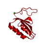 4tnoC  4wabC  4wbqC 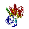 4wbxC C: citing same article ( |
|---|---|
| Similar structure data |


-Links
 PDBj
PDBj


- Assembly
| Deposited unit | 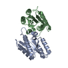
| ||||||||
|---|---|---|---|---|---|---|---|---|---|
| 1 | 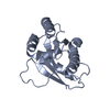
| ||||||||
| 2 | 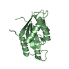
| ||||||||
| Unit cell |
|
-Components
| #1: Protein | Mass: 19245.443 Da / Num. of mol.: 2 Source method: isolated from a genetically manipulated source Details: residue 62 can not be observed in the density, however the density for the whole loop region is not very good. Source: (gene. exp.) Homo sapiens (human) / Gene: CENPM, C22orf18, ICEN39, PANE1 / Production host:  #2: Water | ChemComp-HOH / |  Mass: 18.015 Da / Num. of mol.: 341 / Source method: isolated from a natural source / Formula: H2O Mass: 18.015 Da / Num. of mol.: 341 / Source method: isolated from a natural source / Formula: H2O |
|---|
-Experimental details
-Experiment
| Experiment | Method: X-RAY DIFFRACTION |
|---|
- Sample preparation
| Crystal | Density Matthews: 2.75 Å3/Da / Density % sol: 55.34 % |
|---|---|
| Crystal grow | Temperature: 293.15 K / Method: vapor diffusion, hanging drop / Details: 100 mM bicine pH 8.5, 11 % MPD, 8 mM spermidine / PH range: 8.5 |
-Data collection
| Diffraction | Mean temperature: 100 K |
|---|---|
| Diffraction source | Source: SYNCHROTRON / Site: SLS  / Beamline: X06DA / Wavelength: 2.0664 Å / Beamline: X06DA / Wavelength: 2.0664 Å |
| Detector | Type: DECTRIS PILATUS 2M-F / Detector: PIXEL / Date: Dec 10, 2012 |
| Radiation | Protocol: SINGLE WAVELENGTH / Monochromatic (M) / Laue (L): M / Scattering type: x-ray |
| Radiation wavelength | Wavelength: 2.0664 Å / Relative weight: 1 |
| Reflection | Resolution: 2.2→50 Å / Num. obs: 40969 / % possible obs: 98.1 % / Redundancy: 140.4 % / Net I/σ(I): 92.37 |
| Reflection shell | Resolution: 2.2→2.26 Å / Redundancy: 32.3 % / Rmerge(I) obs: 0.139 / Mean I/σ(I) obs: 16.94 / % possible all: 86.8 |
- Processing
| Software |
| ||||||||||||||||||||||||||||||||||||||||||||||||||||||||||||||||||||||||||||||||||||||||||||||||||||||||||||||||
|---|---|---|---|---|---|---|---|---|---|---|---|---|---|---|---|---|---|---|---|---|---|---|---|---|---|---|---|---|---|---|---|---|---|---|---|---|---|---|---|---|---|---|---|---|---|---|---|---|---|---|---|---|---|---|---|---|---|---|---|---|---|---|---|---|---|---|---|---|---|---|---|---|---|---|---|---|---|---|---|---|---|---|---|---|---|---|---|---|---|---|---|---|---|---|---|---|---|---|---|---|---|---|---|---|---|---|---|---|---|---|---|---|---|
| Refinement | Method to determine structure: SAD / Resolution: 2.2→45.28 Å / Cross valid method: FREE R-VALUE / σ(F): 1.7 / Phase error: 14.5 / Stereochemistry target values: TWIN_LSQ_F
| ||||||||||||||||||||||||||||||||||||||||||||||||||||||||||||||||||||||||||||||||||||||||||||||||||||||||||||||||
| Solvent computation | Shrinkage radii: 0.9 Å / VDW probe radii: 1.11 Å / Solvent model: FLAT BULK SOLVENT MODEL | ||||||||||||||||||||||||||||||||||||||||||||||||||||||||||||||||||||||||||||||||||||||||||||||||||||||||||||||||
| Refinement step | Cycle: LAST / Resolution: 2.2→45.28 Å
| ||||||||||||||||||||||||||||||||||||||||||||||||||||||||||||||||||||||||||||||||||||||||||||||||||||||||||||||||
| Refine LS restraints |
| ||||||||||||||||||||||||||||||||||||||||||||||||||||||||||||||||||||||||||||||||||||||||||||||||||||||||||||||||
| LS refinement shell |
|
