 Movie
Movie Controller

[English] 日本語
 Yorodumi
- PDB-4w6z: YEAST ALCOHOL DEHYDROGENASE I, SACCHAROMYCES CEREVISIAE FERMENTAT... -
+ Open data

- Basic information
| Entry | Database: PDB / ID: 4w6z | |||||||||
|---|---|---|---|---|---|---|---|---|---|---|
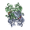
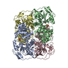
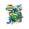
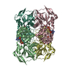
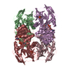
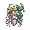
| Title | YEAST ALCOHOL DEHYDROGENASE I, SACCHAROMYCES CEREVISIAE FERMENTATIVE ENZYME | |||||||||
 Components Components | Alcohol dehydrogenase 1 | |||||||||
 Keywords Keywords | OXIDOREDUCTASE / TETRAMER OF ASYMMETRIC DIMERS / ZINC COORDINATION / INTRAMOLECULAR DISULFIDE BONDS | |||||||||
| Function / homology |  Function and homology information Function and homology informationmethylglyoxal reductase (NADH) / octanol dehydrogenase (NAD+) activity / methylglyoxal reductase (NADH) activity / pyruvate fermentation to ethanol / : / butanol dehydrogenase (NAD+) activity / ethanol dehydrogenase (NAD+) activity / melatonin binding / alcohol dehydrogenase (NAD+) activity / allyl-alcohol dehydrogenase ...methylglyoxal reductase (NADH) / octanol dehydrogenase (NAD+) activity / methylglyoxal reductase (NADH) activity / pyruvate fermentation to ethanol / : / butanol dehydrogenase (NAD+) activity / ethanol dehydrogenase (NAD+) activity / melatonin binding / alcohol dehydrogenase (NAD+) activity / allyl-alcohol dehydrogenase / allyl-alcohol dehydrogenase activity / alcohol dehydrogenase / zinc ion binding / identical protein binding / plasma membrane / cytoplasm Similarity search - Function | |||||||||
| Biological species |  | |||||||||
| Method |  X-RAY DIFFRACTION / SYNCHROTRON / MOLECULAR REPLACEMENT / Resolution: 2.4 Å X-RAY DIFFRACTION / SYNCHROTRON / MOLECULAR REPLACEMENT / Resolution: 2.4 Å | |||||||||
 Authors Authors | plapp, B.v. / savarimuthu, b.r. / ramaswamy, s. | |||||||||
| Funding support |  United States, 2items United States, 2items
| |||||||||
 Citation Citation | Journal: Biochemistry / Year: 2014 Title: Yeast alcohol dehydrogenase structure and catalysis. Authors: Savarimuthu Baskar Raj / S Ramaswamy / Bryce V Plapp / Abstract: Yeast (Saccharomyces cerevisiae) alcohol dehydrogenase I (ADH1) is the constitutive enzyme that reduces acetaldehyde to ethanol during the fermentation of glucose. ADH1 is a homotetramer of subunits ...Yeast (Saccharomyces cerevisiae) alcohol dehydrogenase I (ADH1) is the constitutive enzyme that reduces acetaldehyde to ethanol during the fermentation of glucose. ADH1 is a homotetramer of subunits with 347 amino acid residues. A structure for ADH1 was determined by X-ray crystallography at 2.4 Å resolution. The asymmetric unit contains four different subunits, arranged as similar dimers named AB and CD. The unit cell contains two different tetramers made up of "back-to-back" dimers, AB:AB and CD:CD. The A and C subunits in each dimer are structurally similar, with a closed conformation, bound coenzyme, and the oxygen of 2,2,2-trifluoroethanol ligated to the catalytic zinc in the classical tetrahedral coordination with Cys-43, Cys-153, and His-66. In contrast, the B and D subunits have an open conformation with no bound coenzyme, and the catalytic zinc has an alternative, inverted coordination with Cys-43, Cys-153, His-66, and the carboxylate of Glu-67. The asymmetry in the dimeric subunits of the tetramer provides two structures that appear to be relevant for the catalytic mechanism. The alternative coordination of the zinc may represent an intermediate in the mechanism of displacement of the zinc-bound water with alcohol or aldehyde substrates. Substitution of Glu-67 with Gln-67 decreases the catalytic efficiency by 100-fold. Previous studies of structural modeling, evolutionary relationships, substrate specificity, chemical modification, and site-directed mutagenesis are interpreted more fully with the three-dimensional structure. #1: Journal: J. Mol. Biol. / Year: 1994 Title: Crystallization and preliminary crystallographic studies of Saccharomyces cerevisiae alcohol dehydrogenase I. Authors: Ramaswamy, S. / Kratzer, D.A. / Hershey, A.D. / Rogers, P.H. / Arnone, A. / Eklund, H. / Plapp, B.V. #2: Journal: J. Biol. Chem. / Year: 1987 Title: Kinetic characterization of yeast alcohol dehydrogenases. Amino acid residue 294 and substrate specificity. Authors: Ganzhorn, A.J. / Green, D.W. / Hershey, A.D. / Gould, R.M. / Plapp, B.V. | |||||||||
| History |
|
- Structure visualization
| Structure viewer | Molecule: MolmilJmol/JSmol |
|---|
- Downloads & links
-Download
| PDBx/mmCIF format | 4w6z.cif.gz | 522.9 KB | Display | PDBx/mmCIF format |
|---|---|---|---|---|
| PDB format | pdb4w6z.ent.gz | 432.1 KB | Display | PDB format |
| PDBx/mmJSON format | 4w6z.json.gz | Tree view | PDBx/mmJSON format | |
| Others |  Other downloads Other downloads |
-Validation report
| Arichive directory | https://data.pdbj.org/pub/pdb/validation_reports/w6/4w6zftp://data.pdbj.org/pub/pdb/validation_reports/w6/4w6z | HTTPS FTP |
|---|
-Related structure data
| Related structure data | 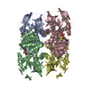 1lluS S: Starting model for refinement |
|---|---|
| Similar structure data |


-Links
 PDBj
PDBj

- Assembly
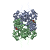
| Deposited unit | 
| ||||||||
|---|---|---|---|---|---|---|---|---|---|
| 1 | 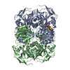
| ||||||||
| 2 | 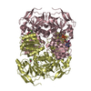
| ||||||||
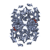
| Unit cell |
| ||||||||
| Components on special symmetry positions |
|
-Components
| #1: Protein | Mass: 36759.906 Da / Num. of mol.: 4 Source method: isolated from a genetically manipulated source Source: (gene. exp.) Strain: ATCC 204508 / S288c / Gene: ADH1, ADC1, YOL086C, O0947 / Plasmid: YEP13 / Production host: Strain (production host): 302-21#2 (ADH1-11, ADH2, LEU2, TRP2) References: UniProt: P00330, alcohol dehydrogenase #2: Chemical | ChemComp-ZN /   Mass: 65.409 Da / Num. of mol.: 8 / Source method: obtained synthetically / Formula: Zn Mass: 65.409 Da / Num. of mol.: 8 / Source method: obtained synthetically / Formula: Zn#3: Chemical |   Mass: 790.330 Da / Num. of mol.: 2 / Source method: obtained synthetically / Formula: C21H27IN7O14P2 Mass: 790.330 Da / Num. of mol.: 2 / Source method: obtained synthetically / Formula: C21H27IN7O14P2#4: Chemical | ChemComp-ETF /   Mass: 100.040 Da / Num. of mol.: 4 / Source method: obtained synthetically / Formula: C2H3F3O Mass: 100.040 Da / Num. of mol.: 4 / Source method: obtained synthetically / Formula: C2H3F3O#5: Water | ChemComp-HOH / |  Mass: 18.015 Da / Num. of mol.: 151 / Source method: isolated from a natural source / Formula: H2O Mass: 18.015 Da / Num. of mol.: 151 / Source method: isolated from a natural source / Formula: H2OHas protein modification | Y | Sequence details | THERE IS AN ERROR IN THE ORIGINAL PUBLISHED DNA SEQUENCE. THE PROTEIN SEQUENCE SHOULD BE TYR AT ...THERE IS AN ERROR IN THE ORIGINAL PUBLISHED DNA SEQUENCE. THE PROTEIN SEQUENCE SHOULD BE TYR AT POSITION 20 (SEE REFERENCE 2 ABOVE). | |
|---|
-Experimental details
-Experiment
| Experiment | Method: X-RAY DIFFRACTION / Number of used crystals: 1 |
|---|
- Sample preparation
| Crystal | Density Matthews: 2.62 Å3/Da / Density % sol: 53.07 % / Description: plate |
|---|---|
| Crystal grow | Temperature: 278 K / Method: vapor diffusion, hanging drop / pH: 8.4 Details: 125 MM SODIUM N-TRIS(HYDROXYMETHYL) METHYL-3-AMINOPROPANE SULFONIC ACID, 1.7 MM NICOTINAMIDE 8- IODOADENINE DINUCLEOTIDE, 0.1 M 2,2,2-TRIFLUOROETHANOL, 0.16 MM EDTA, 10 MG/ML PROTEIN, 6% ...Details: 125 MM SODIUM N-TRIS(HYDROXYMETHYL) METHYL-3-AMINOPROPANE SULFONIC ACID, 1.7 MM NICOTINAMIDE 8- IODOADENINE DINUCLEOTIDE, 0.1 M 2,2,2-TRIFLUOROETHANOL, 0.16 MM EDTA, 10 MG/ML PROTEIN, 6% INITIAL POLYETHYLENE GLYCOL 5000 MONOMETHYL ETHER (FLUKA MPEG5000) IN DROP HANGING OVER 22-26% MPEG5000 AND 0.1 M 2,2,2-TRIFLUOROETHANOL. CRYSTALS WERE SOAKED IN SAME BUFFER WITH 30% W/V MPEG5000 WITH 0.5 M 2,2,2- TRIFLUOROETHANOL FOR FIVE DAYS BEFORE FREEZING AT 100 K., PH 8.4, VAPOR DIFFUSION, HANGING DROP, TEMPERATURE 278K, PH 8.40 PH range: 8.4 |
-Data collection
| Diffraction | Mean temperature: 100 K | |||||||||||||||
|---|---|---|---|---|---|---|---|---|---|---|---|---|---|---|---|---|
| Diffraction source | Source: SYNCHROTRON / Site: EMBL/DESY, HAMBURG  / Beamline: BW7A / Wavelength: 0.857 Å / Beamline: BW7A / Wavelength: 0.857 Å | |||||||||||||||
| Detector | Type: MAR scanner 300 mm plate / Detector: IMAGE PLATE / Date: Jul 28, 1995 / Details: MULTIPOLE WIGGLER | |||||||||||||||
| Radiation | Protocol: SINGLE WAVELENGTH / Monochromatic (M) / Laue (L): M / Scattering type: x-ray | |||||||||||||||
| Radiation wavelength | Wavelength: 0.857 Å / Relative weight: 1 | |||||||||||||||
| Reflection twin |
| |||||||||||||||
| Reflection | Resolution: 2.4→40.4 Å / Num. obs: 59662 / % possible obs: 98.5 % / Redundancy: 3.85 % / Rmerge(I) obs: 0.116 / Rsym value: 0.116 / Net I/σ(I): 8 | |||||||||||||||
| Reflection shell | Resolution: 2.4→2.49 Å / Redundancy: 3.71 % / Rmerge(I) obs: 0.418 / Mean I/σ(I) obs: 3.5 / Rsym value: 0.418 / % possible all: 93.3 |
- Processing
| Software |
| ||||||||||||||||||||||||||||||||||||||||||||||||||||||||||||||||||||||||||||||||||||||||||||||||||||||||||||||||||||||||||||||||||||||||||||||||||||||||||||||||||||||||||||||||||||||
|---|---|---|---|---|---|---|---|---|---|---|---|---|---|---|---|---|---|---|---|---|---|---|---|---|---|---|---|---|---|---|---|---|---|---|---|---|---|---|---|---|---|---|---|---|---|---|---|---|---|---|---|---|---|---|---|---|---|---|---|---|---|---|---|---|---|---|---|---|---|---|---|---|---|---|---|---|---|---|---|---|---|---|---|---|---|---|---|---|---|---|---|---|---|---|---|---|---|---|---|---|---|---|---|---|---|---|---|---|---|---|---|---|---|---|---|---|---|---|---|---|---|---|---|---|---|---|---|---|---|---|---|---|---|---|---|---|---|---|---|---|---|---|---|---|---|---|---|---|---|---|---|---|---|---|---|---|---|---|---|---|---|---|---|---|---|---|---|---|---|---|---|---|---|---|---|---|---|---|---|---|---|---|---|
| Refinement | Method to determine structure: MOLECULAR REPLACEMENT Starting model: 1LLU.PDB Resolution: 2.4→28.09 Å / Cor.coef. Fo:Fc: 0.945 / Cor.coef. Fo:Fc free: 0.915 / SU B: 11.881 / SU ML: 0.15 / Cross valid method: THROUGHOUT / ESU R: 0.075 / ESU R Free: 0.049 / Stereochemistry target values: MAXIMUM LIKELIHOOD / Details: HYDROGENS HAVE BEEN ADDED IN THE RIDING POSITIONS
| ||||||||||||||||||||||||||||||||||||||||||||||||||||||||||||||||||||||||||||||||||||||||||||||||||||||||||||||||||||||||||||||||||||||||||||||||||||||||||||||||||||||||||||||||||||||
| Solvent computation | Ion probe radii: 0.8 Å / Shrinkage radii: 0.8 Å / VDW probe radii: 1.2 Å / Solvent model: MASK | ||||||||||||||||||||||||||||||||||||||||||||||||||||||||||||||||||||||||||||||||||||||||||||||||||||||||||||||||||||||||||||||||||||||||||||||||||||||||||||||||||||||||||||||||||||||
| Displacement parameters | Biso mean: 40.195 Å2
| ||||||||||||||||||||||||||||||||||||||||||||||||||||||||||||||||||||||||||||||||||||||||||||||||||||||||||||||||||||||||||||||||||||||||||||||||||||||||||||||||||||||||||||||||||||||
| Refinement step | Cycle: 1 / Resolution: 2.4→28.09 Å
| ||||||||||||||||||||||||||||||||||||||||||||||||||||||||||||||||||||||||||||||||||||||||||||||||||||||||||||||||||||||||||||||||||||||||||||||||||||||||||||||||||||||||||||||||||||||
| Refine LS restraints |
|