 Movie
Movie Controller


+ Open data

- Basic information
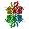
| Entry | Database: PDB / ID: 1pl8 | ||||||
|---|---|---|---|---|---|---|---|
| Title | human SDH/NAD+ complex | ||||||
 Components Components | human sorbitol dehydrogenase | ||||||
 Keywords Keywords | OXIDOREDUCTASE / human sorbitol dehydrogenase / NAD | ||||||
| Function / homology |  Function and homology information Function and homology informationribitol 2-dehydrogenase / ribitol 2-dehydrogenase (NAD+) activity / xylitol catabolic process / xylitol metabolic process / (R,R)-butanediol dehydrogenase / (R,R)-butanediol dehydrogenase activity / D-xylulose reductase / D-xylulose reductase activity / L-iditol 2-dehydrogenase / D-sorbitol catabolic process ...ribitol 2-dehydrogenase / ribitol 2-dehydrogenase (NAD+) activity / xylitol catabolic process / xylitol metabolic process / (R,R)-butanediol dehydrogenase / (R,R)-butanediol dehydrogenase activity / D-xylulose reductase / D-xylulose reductase activity / L-iditol 2-dehydrogenase / D-sorbitol catabolic process / L-iditol 2-dehydrogenase (NAD+) activity / Formation of xylulose-5-phosphate / : / Fructose biosynthesis / fructose biosynthetic process / flagellated sperm motility / Oxidoreductases; Acting on the CH-OH group of donors; With NAD+ or NADP+ as acceptor / motile cilium / glucose metabolic process / mitochondrial membrane / NAD binding / carbohydrate binding / : / extracellular exosome / zinc ion binding / membrane / identical protein binding / cytosol Similarity search - Function | ||||||
| Biological species |  Homo sapiens (human) Homo sapiens (human) | ||||||
| Method |  X-RAY DIFFRACTION / SYNCHROTRON / MOLECULAR REPLACEMENT / Resolution: 1.9 Å X-RAY DIFFRACTION / SYNCHROTRON / MOLECULAR REPLACEMENT / Resolution: 1.9 Å | ||||||
 Authors Authors | Pauly, T.A. / Ekstrom, J.L. / Beebe, D.A. / Chrunyk, B. / Cunningham, D. / Griffor, M. / Kamath, A. / Lee, S.E. / Madura, R. / Mcguire, D. ...Pauly, T.A. / Ekstrom, J.L. / Beebe, D.A. / Chrunyk, B. / Cunningham, D. / Griffor, M. / Kamath, A. / Lee, S.E. / Madura, R. / Mcguire, D. / Subashi, T. / Wasilko, D. / Watts, P. / Mylari, B.L. / Oates, P.J. / Adams, P.D. / Rath, V.L. | ||||||
 Citation Citation | Journal: Structure / Year: 2003 Title: X-ray crystallographic and kinetic studies of human sorbitol dehydrogenase. Authors: Pauly, T.A. / Ekstrom, J.L. / Beebe, D.A. / Chrunyk, B. / Cunningham, D. / Griffor, M. / Kamath, A. / Lee, S.E. / Madura, R. / Mcguire, D. / Subashi, T. / Wasilko, D. / Watts, P. / Mylari, B. ...Authors: Pauly, T.A. / Ekstrom, J.L. / Beebe, D.A. / Chrunyk, B. / Cunningham, D. / Griffor, M. / Kamath, A. / Lee, S.E. / Madura, R. / Mcguire, D. / Subashi, T. / Wasilko, D. / Watts, P. / Mylari, B.L. / Oates, P.J. / Adams, P.D. / Rath, V.L. | ||||||
| History |
| ||||||
| Remark 999 | The sequence variant Leu -> Gln for residue 238 is noted in Swiss-Prot entry Q00796. |
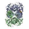
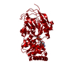
- Structure visualization
| Structure viewer | Molecule: MolmilJmol/JSmol |
|---|
- Downloads & links
-Download
| PDBx/mmCIF format | 1pl8.cif.gz | 308.8 KB | Display | PDBx/mmCIF format |
|---|---|---|---|---|
| PDB format | pdb1pl8.ent.gz | 249.8 KB | Display | PDB format |
| PDBx/mmJSON format | 1pl8.json.gz | Tree view | PDBx/mmJSON format | |
| Others |  Other downloads Other downloads |
-Validation report
| Arichive directory | https://data.pdbj.org/pub/pdb/validation_reports/pl/1pl8ftp://data.pdbj.org/pub/pdb/validation_reports/pl/1pl8 | HTTPS FTP |
|---|
-Related structure data


-Links
 PDBj
PDBj

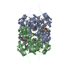
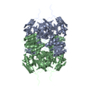
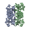
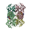
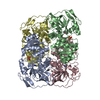
- Assembly
| Deposited unit | 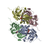
| ||||||||
|---|---|---|---|---|---|---|---|---|---|
| 1 | 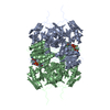
| ||||||||
| 2 | 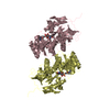
| ||||||||
| 3 |
| ||||||||
| Unit cell |
|
-Components
| #1: Protein | Mass: 38229.332 Da / Num. of mol.: 4 Source method: isolated from a genetically manipulated source Source: (gene. exp.) Homo sapiens (human) / Species (production host): Escherichia coli / Production host:  #2: Chemical | ChemComp-ZN /   Mass: 65.409 Da / Num. of mol.: 4 / Source method: obtained synthetically / Formula: Zn Mass: 65.409 Da / Num. of mol.: 4 / Source method: obtained synthetically / Formula: Zn#3: Chemical | ChemComp-NAD /   Mass: 663.425 Da / Num. of mol.: 4 / Source method: obtained synthetically / Formula: C21H27N7O14P2 / Comment: NAD*YM Mass: 663.425 Da / Num. of mol.: 4 / Source method: obtained synthetically / Formula: C21H27N7O14P2 / Comment: NAD*YM#4: Water | ChemComp-HOH / |  Mass: 18.015 Da / Num. of mol.: 1291 / Source method: isolated from a natural source / Formula: H2O Mass: 18.015 Da / Num. of mol.: 1291 / Source method: isolated from a natural source / Formula: H2O |
|---|
-Experimental details
-Experiment
| Experiment | Method: X-RAY DIFFRACTION / Number of used crystals: 1 |
|---|
- Sample preparation
| Crystal | Density Matthews: 3.79 Å3/Da / Density % sol: 67.57 % | ||||||||||||||||||||||||||||||||||||
|---|---|---|---|---|---|---|---|---|---|---|---|---|---|---|---|---|---|---|---|---|---|---|---|---|---|---|---|---|---|---|---|---|---|---|---|---|---|
| Crystal grow | Temperature: 295 K / Method: vapor diffusion, hanging drop / pH: 6.15 Details: NH4AC, NaCitrate, PEG 4000, pH 6.15, VAPOR DIFFUSION, HANGING DROP, temperature 295K | ||||||||||||||||||||||||||||||||||||
| Crystal grow | *PLUS Method: vapor diffusion | ||||||||||||||||||||||||||||||||||||
| Components of the solutions | *PLUS
|
-Data collection
| Diffraction | Mean temperature: 100 K |
|---|---|
| Diffraction source | Source: SYNCHROTRON / Site: NSLS  / Beamline: X12C / Wavelength: 1 Å / Beamline: X12C / Wavelength: 1 Å |
| Detector | Type: BRANDEIS - B1.2 / Detector: CCD / Date: May 18, 2001 Details: double-crystal monochromator Si(111), beam focused by a toroidal mirror |
| Radiation | Monochromator: double-crystal monochromator Si(111) / Protocol: SINGLE WAVELENGTH / Monochromatic (M) / Laue (L): M / Scattering type: x-ray |
| Radiation wavelength | Wavelength: 1 Å / Relative weight: 1 |
| Reflection | Resolution: 1.9→99 Å / Num. obs: 161025 / % possible obs: 88.6 % / Observed criterion σ(I): 2 / Redundancy: 3.2 % / Biso Wilson estimate: 11.8 Å2 / Rsym value: 0.083 / Net I/σ(I): 12.8 |
| Reflection shell | Resolution: 1.9→1.97 Å / Redundancy: 1 % / Rmerge(I) obs: 0.379 / Mean I/σ(I) obs: 2 / Num. unique all: 7993 / % possible all: 44.2 |
| Reflection | *PLUS Highest resolution: 1.9 Å / Redundancy: 4 % / Rmerge(I) obs: 0.083 |
| Reflection shell | *PLUS % possible obs: 44.2 % |
- Processing
| Software |
| ||||||||||||||||||||||||||||||||||||||||||||||||||||||||||||
|---|---|---|---|---|---|---|---|---|---|---|---|---|---|---|---|---|---|---|---|---|---|---|---|---|---|---|---|---|---|---|---|---|---|---|---|---|---|---|---|---|---|---|---|---|---|---|---|---|---|---|---|---|---|---|---|---|---|---|---|---|---|
| Refinement | Method to determine structure: MOLECULAR REPLACEMENT / Resolution: 1.9→45.82 Å / Rfactor Rfree error: 0.002 / Isotropic thermal model: RESTRAINED / Cross valid method: THROUGHOUT / σ(F): 0 / Stereochemistry target values: Engh & Huber
| ||||||||||||||||||||||||||||||||||||||||||||||||||||||||||||
| Solvent computation | Solvent model: FLAT MODEL / Bsol: 54.2901 Å2 / ksol: 0.401316 e/Å3 | ||||||||||||||||||||||||||||||||||||||||||||||||||||||||||||
| Displacement parameters | Biso mean: 23.2 Å2
| ||||||||||||||||||||||||||||||||||||||||||||||||||||||||||||
| Refine analyze | Luzzati coordinate error free: 0.25 Å / Luzzati sigma a free: 0.24 Å | ||||||||||||||||||||||||||||||||||||||||||||||||||||||||||||
| Refinement step | Cycle: LAST / Resolution: 1.9→45.82 Å
| ||||||||||||||||||||||||||||||||||||||||||||||||||||||||||||
| Refine LS restraints |
| ||||||||||||||||||||||||||||||||||||||||||||||||||||||||||||
| LS refinement shell | Resolution: 1.9→2.02 Å / Rfactor Rfree error: 0.008 / Total num. of bins used: 6
| ||||||||||||||||||||||||||||||||||||||||||||||||||||||||||||
| Xplor file |
| ||||||||||||||||||||||||||||||||||||||||||||||||||||||||||||
| Refinement | *PLUS Highest resolution: 1.9 Å / Lowest resolution: 45.8 Å / % reflection Rfree: 10 % | ||||||||||||||||||||||||||||||||||||||||||||||||||||||||||||
| Solvent computation | *PLUS | ||||||||||||||||||||||||||||||||||||||||||||||||||||||||||||
| Displacement parameters | *PLUS | ||||||||||||||||||||||||||||||||||||||||||||||||||||||||||||
| Refine LS restraints | *PLUS
| ||||||||||||||||||||||||||||||||||||||||||||||||||||||||||||
| LS refinement shell | *PLUS Rfactor Rwork: 0.27 |
