 Movie
Movie Controller


+ Open data

- Basic information
| Entry | Database: PDB / ID: 7kjy | ||||||
|---|---|---|---|---|---|---|---|
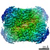
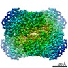
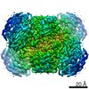
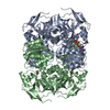
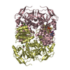
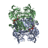
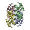
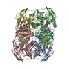
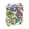
| Title | Symmetry in Yeast Alcohol Dehydrogenase 1 - Open Form with NADH | ||||||
 Components Components | Alcohol dehydrogenase | ||||||
 Keywords Keywords | OXIDOREDUCTASE / Alcohol dehydrogenase / NADH | ||||||
| Function / homology |  Function and homology information Function and homology information: / alcohol dehydrogenase (NAD+) activity / alcohol dehydrogenase / zinc ion binding / cytoplasm Similarity search - Function | ||||||
| Biological species |  | ||||||
| Method | ELECTRON MICROSCOPY / single particle reconstruction / cryo EM / Resolution: 3.2 Å | ||||||
 Authors Authors | Subramanian, R. / Chang, L. / Li, Z. / Plapp, B.V. / Guntupalli, S.R. | ||||||
 Citation Citation | Journal: Biochemistry / Year: 2021 Title: Cryo-Electron Microscopy Structures of Yeast Alcohol Dehydrogenase. Authors: Sai Rohit Guntupalli / Zhuang Li / Leifu Chang / Bryce V Plapp / Ramaswamy Subramanian /   Abstract: Structures of yeast alcohol dehydrogenase determined by X-ray crystallography show that the subunits have two different conformational states in each of the two dimers that form the tetramer. ...Structures of yeast alcohol dehydrogenase determined by X-ray crystallography show that the subunits have two different conformational states in each of the two dimers that form the tetramer. Apoenzyme and holoenzyme complexes relevant to the catalytic mechanism were described, but the asymmetry led to questions about the cooperativity of the subunits in catalysis. This study used cryo-electron microscopy (cryo-EM) to provide structures for the apoenzyme, two different binary complexes with NADH, and a ternary complex with NAD and 2,2,2-trifluoroethanol. All four subunits in each of these complexes are identical, as the tetramers have 2 symmetry, suggesting that there is no preexisting asymmetry and that the subunits can be independently active. The apoenzyme and one enzyme-NADH complex have "open" conformations and the inverted coordination of the catalytic zinc with Cys-43, His-66, Glu-67, and Cys-153, whereas another enzyme-NADH complex and the ternary complex have closed conformations with the classical coordination of the zinc with Cys-43, His-66, Cys-153, and a water or the oxygen of trifluoroethanol. The conformational change involves interactions of Arg-340 with the pyrophosphate group of the coenzyme and Glu-67. The cryo-EM and X-ray crystallography studies provide structures relevant for the catalytic mechanism. #1: Journal: Arch Biochem Biophys / Year: 2016Title: Mechanistic implications from structures of yeast alcohol dehydrogenase complexed with coenzyme and an alcohol. Authors: Plapp, B.V. / Charlier, H.A. / Ramaswamy, S. | ||||||
| History |
|
- Structure visualization
| Movie |
Movie viewer |
|---|---|
| Structure viewer | Molecule: MolmilJmol/JSmol |
- Downloads & links
-Download
| PDBx/mmCIF format | 7kjy.cif.gz | 237.4 KB | Display | PDBx/mmCIF format |
|---|---|---|---|---|
| PDB format | pdb7kjy.ent.gz | 191.7 KB | Display | PDB format |
| PDBx/mmJSON format | 7kjy.json.gz | Tree view | PDBx/mmJSON format | |
| Others |  Other downloads Other downloads |
-Validation report
| Arichive directory | https://data.pdbj.org/pub/pdb/validation_reports/kj/7kjyftp://data.pdbj.org/pub/pdb/validation_reports/kj/7kjy | HTTPS FTP |
|---|
-Related structure data
| Related structure data | 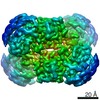 22902MC 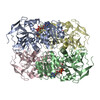 7kc2C 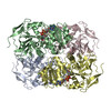 7kcbC 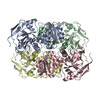 7kcqC C: citing same article ( M: map data used to model this data |
|---|---|
| Similar structure data |


-Links
 PDBj
PDBj

- Assembly
| Deposited unit | 
|
|---|---|
| 1 |
|
-Components
| #1: Protein | Mass: 36881.016 Da / Num. of mol.: 4 / Source method: isolated from a natural source / Source: (natural) #2: Chemical | ChemComp-ZN /   Mass: 65.409 Da / Num. of mol.: 8 / Source method: obtained synthetically / Formula: Zn / Feature type: SUBJECT OF INVESTIGATION Mass: 65.409 Da / Num. of mol.: 8 / Source method: obtained synthetically / Formula: Zn / Feature type: SUBJECT OF INVESTIGATION#3: Chemical | ChemComp-NAD /   Mass: 663.425 Da / Num. of mol.: 4 / Source method: obtained synthetically / Formula: C21H27N7O14P2 / Feature type: SUBJECT OF INVESTIGATION / Comment: NAD*YM Mass: 663.425 Da / Num. of mol.: 4 / Source method: obtained synthetically / Formula: C21H27N7O14P2 / Feature type: SUBJECT OF INVESTIGATION / Comment: NAD*YMHas ligand of interest | Y | Has protein modification | Y | |
|---|
-Experimental details
-Experiment
| Experiment | Method: ELECTRON MICROSCOPY |
|---|---|
| EM experiment | Aggregation state: PARTICLE / 3D reconstruction method: single particle reconstruction |
- Sample preparation
| Component | Name: Sc Alcohol Dehydrogenase NADH Complex / Type: COMPLEX / Entity ID: #1 / Source: NATURAL |
|---|---|
| Molecular weight | Value: 0.37 MDa / Experimental value: NO |
| Source (natural) | Organism: |
| Buffer solution | pH: 8.2 Details: Tris HCl buffer 5mM with 200mM KCl adjusted to pH 8.2. |
| Buffer component | Conc.: 50 mM / Name: Tris / Formula: C4H11NO3 |
| Specimen | Conc.: 5 mg/ml / Embedding applied: NO / Shadowing applied: NO / Staining applied: NO / Vitrification applied: YES / Details: Purified by Size Exclusion chromatography |
| Specimen support | Details: unspecified |
| Vitrification | Instrument: FEI VITROBOT MARK II / Cryogen name: ETHANE / Humidity: 100 % / Chamber temperature: 298 K |
- Electron microscopy imaging
| Experimental equipment |  Model: Titan Krios / Image courtesy: FEI Company |
|---|---|
| Microscopy | Model: FEI TITAN KRIOS |
| Electron gun | Electron source:  FIELD EMISSION GUN / Accelerating voltage: 300 kV / Illumination mode: OTHER FIELD EMISSION GUN / Accelerating voltage: 300 kV / Illumination mode: OTHER |
| Electron lens | Mode: BRIGHT FIELD / Nominal defocus max: 2000 nm / Nominal defocus min: 800 nm / Cs: 2.7 mm / C2 aperture diameter: 100 µm |
| Specimen holder | Cryogen: NITROGEN |
| Image recording | Electron dose: 54 e/Å2 / Film or detector model: GATAN K3 BIOQUANTUM (6k x 4k) |
- Processing
| Software | Name: PHENIX / Version: 1.17.1_3660: / Classification: refinement | ||||||||||||||||||||||||
|---|---|---|---|---|---|---|---|---|---|---|---|---|---|---|---|---|---|---|---|---|---|---|---|---|---|
| EM software |
| ||||||||||||||||||||||||
| CTF correction | Type: NONE | ||||||||||||||||||||||||
| Symmetry | Point symmetry: D2 (2x2 fold dihedral) | ||||||||||||||||||||||||
| 3D reconstruction | Resolution: 3.2 Å / Resolution method: FSC 0.143 CUT-OFF / Num. of particles: 476561 / Symmetry type: POINT | ||||||||||||||||||||||||
| Atomic model building | B value: 39.3 / Protocol: RIGID BODY FIT / Space: REAL / Target criteria: Correlation Coefficient | ||||||||||||||||||||||||
| Atomic model building | PDB-ID: 4W6Z Pdb chain-ID: B / Accession code: 4W6Z / Pdb chain residue range: 1-347 / Source name: PDB / Type: experimental model | ||||||||||||||||||||||||
| Refine LS restraints |
|
