 Movie
Movie Controller

[English] 日本語
 Yorodumi
- PDB-1llu: THE TERNARY COMPLEX OF PSEUDOMONAS AERUGINOSA ALCOHOL DEHYDROGENA... -
+ Open data

- Basic information
| Entry | Database: PDB / ID: 1llu | ||||||
|---|---|---|---|---|---|---|---|
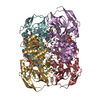
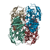
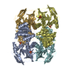
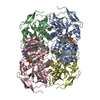
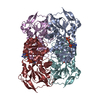
| Title | THE TERNARY COMPLEX OF PSEUDOMONAS AERUGINOSA ALCOHOL DEHYDROGENASE WITH ITS COENZYME AND WEAK SUBSTRATE | ||||||
 Components Components | Alcohol Dehydrogenase | ||||||
 Keywords Keywords | OXIDOREDUCTASE / Enzyme-coenzyme-substrate complex / proton relay system / NADH / Alcohol Dehydrogenase | ||||||
| Function / homology |  Function and homology information Function and homology informationalcohol dehydrogenase (NAD+) activity / alcohol dehydrogenase / nucleotide binding / zinc ion binding Similarity search - Function | ||||||
| Biological species |   Pseudomonas aeruginosa (bacteria) Pseudomonas aeruginosa (bacteria) | ||||||
| Method |  X-RAY DIFFRACTION / MOLECULAR REPLACEMENT / Resolution: 2.3 Å X-RAY DIFFRACTION / MOLECULAR REPLACEMENT / Resolution: 2.3 Å | ||||||
 Authors Authors | Levin, I. / Meiri, G. / Peretz, M. / Frolow, F. / Burstein, Y. | ||||||
 Citation Citation | Journal: Protein Sci. / Year: 2004 Title: The ternary complex of Pseudomonas aeruginosa alcohol dehydrogenase with NADH and ethylene glycol. Authors: Levin, I. / Meiri, G. / Peretz, M. / Burstein, Y. / Frolow, F. | ||||||
| History |
| ||||||
| Remark 999 | SEQUENCE Author states residue ALA 229 and ILE 230 were verified by DNA sequencing, there are two ...SEQUENCE Author states residue ALA 229 and ILE 230 were verified by DNA sequencing, there are two conflicts between the strain Habs serotype I used in this study (Kessler, E., Safrin, M., Peretz, M., and Burstein, Y. (1992). Identification of cleavage sites involved in proteolytic processing of Pseudomonas aeruginosa preproelastase, FEBS Lett 299, 291-3) and the sequence in data base. |
- Structure visualization
| Structure viewer | Molecule: MolmilJmol/JSmol |
|---|
- Downloads & links
-Download
| PDBx/mmCIF format | 1llu.cif.gz | 529.8 KB | Display | PDBx/mmCIF format |
|---|---|---|---|---|
| PDB format | pdb1llu.ent.gz | 435.2 KB | Display | PDB format |
| PDBx/mmJSON format | 1llu.json.gz | Tree view | PDBx/mmJSON format | |
| Others |  Other downloads Other downloads |
-Validation report
| Arichive directory | https://data.pdbj.org/pub/pdb/validation_reports/ll/1lluftp://data.pdbj.org/pub/pdb/validation_reports/ll/1llu | HTTPS FTP |
|---|
-Related structure data
| Related structure data | 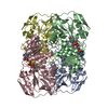 1ykfS S: Starting model for refinement |
|---|---|
| Similar structure data |

-Links
 PDBj
PDBj

- Assembly
| Deposited unit | 
| ||||||||||||
|---|---|---|---|---|---|---|---|---|---|---|---|---|---|
| 1 | 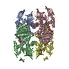
| ||||||||||||
| 2 | 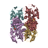
| ||||||||||||
| Unit cell |
| ||||||||||||
| Noncrystallographic symmetry (NCS) | NCS oper:
| ||||||||||||
| Details | Coordinates for biologically active tetramer are provided. The second tetramer in assymetric unit can be generated by rotation: (-0.72417 -0.09256 -0.68339 ), ( -0.09185 -0.96918 0.22859 ), (-0.68348 0.22831 0.69335 ) and translation (in A) (82.48212 18.86833 -11.46263) |
-Components
| #1: Protein | Mass: 35919.180 Da / Num. of mol.: 8 Source method: isolated from a genetically manipulated source Source: (gene. exp.) Pseudomonas aeruginosa (bacteria) / Strain: Habs serotype I / Gene: adh / Plasmid: BS-p80 / Production host: #2: Chemical | ChemComp-ZN /   Mass: 65.409 Da / Num. of mol.: 16 / Source method: obtained synthetically / Formula: Zn Mass: 65.409 Da / Num. of mol.: 16 / Source method: obtained synthetically / Formula: Zn#3: Chemical | ChemComp-NAD /   Mass: 663.425 Da / Num. of mol.: 8 / Source method: obtained synthetically / Formula: C21H27N7O14P2 / Comment: NAD*YM Mass: 663.425 Da / Num. of mol.: 8 / Source method: obtained synthetically / Formula: C21H27N7O14P2 / Comment: NAD*YM#4: Chemical | ChemComp-EDO /   Mass: 62.068 Da / Num. of mol.: 12 / Source method: obtained synthetically / Formula: C2H6O2 Mass: 62.068 Da / Num. of mol.: 12 / Source method: obtained synthetically / Formula: C2H6O2#5: Water | ChemComp-HOH / |  Mass: 18.015 Da / Num. of mol.: 1044 / Source method: isolated from a natural source / Formula: H2O Mass: 18.015 Da / Num. of mol.: 1044 / Source method: isolated from a natural source / Formula: H2O |
|---|
-Experimental details
-Experiment
| Experiment | Method: X-RAY DIFFRACTION / Number of used crystals: 1 |
|---|
- Sample preparation
| Crystal | Density Matthews: 2.58 Å3/Da / Density % sol: 52.35 % | ||||||||||||||||||||||||||||||||||||||||||||||||
|---|---|---|---|---|---|---|---|---|---|---|---|---|---|---|---|---|---|---|---|---|---|---|---|---|---|---|---|---|---|---|---|---|---|---|---|---|---|---|---|---|---|---|---|---|---|---|---|---|---|
| Crystal grow | Temperature: 292 K / Method: vapor diffusion, hanging drop / pH: 8.6 Details: 2mM NAD+, 16mM Spermine, 14% Polyethelene Glycol Monomethyl Ether 2000, 12mM Sodium Potassium Tartrate, 100mM Tris pH 8.6, 0.1mM DTT, 0.007% Sodium Azide, VAPOR DIFFUSION, HANGING DROP, temperature 292.0K | ||||||||||||||||||||||||||||||||||||||||||||||||
| Crystal grow | *PLUS Temperature: 19 K / Method: vapor diffusion, hanging drop | ||||||||||||||||||||||||||||||||||||||||||||||||
| Components of the solutions | *PLUS
|
-Data collection
| Diffraction | Mean temperature: 120 K |
|---|---|
| Diffraction source | Source: ROTATING ANODE / Type: RIGAKU / Wavelength: 1.5418 Å |
| Detector | Type: RIGAKU RAXIS IV++ / Detector: IMAGE PLATE / Date: Nov 27, 2001 / Details: Osmic-Blue confocal optics |
| Radiation | Monochromator: Osmic Mirrors / Protocol: SINGLE WAVELENGTH / Monochromatic (M) / Laue (L): M / Scattering type: x-ray |
| Radiation wavelength | Wavelength: 1.5418 Å / Relative weight: 1 |
| Reflection | Resolution: 2.3→20 Å / Num. all: 123136 / Num. obs: 123136 / % possible obs: 96.4 % / Observed criterion σ(F): 0 / Observed criterion σ(I): -3 / Redundancy: 3.4 % / Biso Wilson estimate: 19.5 Å2 / Rsym value: 0.108 / Net I/σ(I): 12.1 |
| Reflection shell | Resolution: 2.3→2.34 Å / Redundancy: 3.3 % / Mean I/σ(I) obs: 2.8 / Num. unique all: 6019 / Rsym value: 0.463 / % possible all: 94.2 |
| Reflection | *PLUS Highest resolution: 2.3 Å / Rmerge(I) obs: 0.108 |
| Reflection shell | *PLUS Highest resolution: 2.3 Å / % possible obs: 94.2 % / Rmerge(I) obs: 0.463 |
- Processing
| Software |
| ||||||||||||||||||||||||||||||||||||||||||||||||||||||||||||||||||||||||||||||||
|---|---|---|---|---|---|---|---|---|---|---|---|---|---|---|---|---|---|---|---|---|---|---|---|---|---|---|---|---|---|---|---|---|---|---|---|---|---|---|---|---|---|---|---|---|---|---|---|---|---|---|---|---|---|---|---|---|---|---|---|---|---|---|---|---|---|---|---|---|---|---|---|---|---|---|---|---|---|---|---|---|---|
| Refinement | Method to determine structure: MOLECULAR REPLACEMENT Starting model: PDB ENTRY 1YKF Resolution: 2.3→19.99 Å / Rfactor Rfree error: 0.002 / Data cutoff high absF: 1547341.28 / Data cutoff high rms absF: 1547341.28 / Data cutoff low absF: 0 / Isotropic thermal model: RESTRAINED / Cross valid method: THROUGHOUT / σ(F): 0 / σ(I): -3 / Stereochemistry target values: Engh & Huber
| ||||||||||||||||||||||||||||||||||||||||||||||||||||||||||||||||||||||||||||||||
| Solvent computation | Solvent model: FLAT MODEL / Bsol: 48.711 Å2 / ksol: 0.363236 e/Å3 | ||||||||||||||||||||||||||||||||||||||||||||||||||||||||||||||||||||||||||||||||
| Displacement parameters | Biso mean: 31.3 Å2
| ||||||||||||||||||||||||||||||||||||||||||||||||||||||||||||||||||||||||||||||||
| Refine analyze |
| ||||||||||||||||||||||||||||||||||||||||||||||||||||||||||||||||||||||||||||||||
| Refinement step | Cycle: LAST / Resolution: 2.3→19.99 Å
| ||||||||||||||||||||||||||||||||||||||||||||||||||||||||||||||||||||||||||||||||
| Refine LS restraints |
| ||||||||||||||||||||||||||||||||||||||||||||||||||||||||||||||||||||||||||||||||
| Refine LS restraints NCS | NCS model details: CONSTR | ||||||||||||||||||||||||||||||||||||||||||||||||||||||||||||||||||||||||||||||||
| LS refinement shell | Resolution: 2.3→2.38 Å / Rfactor Rfree error: 0.009 / Total num. of bins used: 10
| ||||||||||||||||||||||||||||||||||||||||||||||||||||||||||||||||||||||||||||||||
| Xplor file |
| ||||||||||||||||||||||||||||||||||||||||||||||||||||||||||||||||||||||||||||||||
| Refinement | *PLUS Highest resolution: 2.3 Å | ||||||||||||||||||||||||||||||||||||||||||||||||||||||||||||||||||||||||||||||||
| Solvent computation | *PLUS | ||||||||||||||||||||||||||||||||||||||||||||||||||||||||||||||||||||||||||||||||
| Displacement parameters | *PLUS | ||||||||||||||||||||||||||||||||||||||||||||||||||||||||||||||||||||||||||||||||
| Refine LS restraints | *PLUS
|
