 Movie
Movie Controller

[English] 日本語
 Yorodumi
- PDB-3wle: Crystal structure of (R)-carbonyl reductase from Candida Parapsil... -
+ Open data

- Basic information
| Entry | Database: PDB / ID: 3wle | ||||||
|---|---|---|---|---|---|---|---|
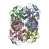
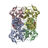
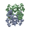
| Title | Crystal structure of (R)-carbonyl reductase from Candida Parapsilosis in complex with NAD | ||||||
 Components Components | (R)-specific carbonyl reductase | ||||||
 Keywords Keywords | OXIDOREDUCTASE / alcohol dehydrogenases / carbonyl reductase | ||||||
| Function / homology |  Function and homology information Function and homology informationalcohol dehydrogenase (NAD+) activity / alcohol dehydrogenase / nucleotide binding / zinc ion binding / cytoplasm Similarity search - Function | ||||||
| Biological species |  Candida parapsilosis (fungus) Candida parapsilosis (fungus) | ||||||
| Method |  X-RAY DIFFRACTION / SYNCHROTRON / SAD / Resolution: 2.164 Å X-RAY DIFFRACTION / SYNCHROTRON / SAD / Resolution: 2.164 Å | ||||||
 Authors Authors | Wang, S.S. / Nie, Y. / Xu, Y. / Zhang, R.Z. / Huang, C.H. / Chan, H.C. / Guo, R.T. / Xiao, R. | ||||||
 Citation Citation | Journal: Chem.Commun.(Camb.) / Year: 2014 Title: Unconserved substrate-binding sites direct the stereoselectivity of medium-chain alcohol dehydrogenase Authors: Wang, S.S. / Nie, Y. / Xu, Y. / Zhang, R.Z. / Ko, T.P. / Huang, C.H. / Chan, H.C. / Guo, R.T. / Xiao, R. | ||||||
| History |
|
- Structure visualization
| Structure viewer | Molecule: MolmilJmol/JSmol |
|---|
- Downloads & links
-Download
| PDBx/mmCIF format | 3wle.cif.gz | 285.3 KB | Display | PDBx/mmCIF format |
|---|---|---|---|---|
| PDB format | pdb3wle.ent.gz | 229.1 KB | Display | PDB format |
| PDBx/mmJSON format | 3wle.json.gz | Tree view | PDBx/mmJSON format | |
| Others |  Other downloads Other downloads |
-Validation report
| Arichive directory | https://data.pdbj.org/pub/pdb/validation_reports/wl/3wleftp://data.pdbj.org/pub/pdb/validation_reports/wl/3wle | HTTPS FTP |
|---|
-Related structure data
| Related structure data |  3wlfC  3wnqC  2xaaS C: citing same article ( S: Starting model for refinement |
|---|---|
| Similar structure data |
-Links
 PDBj
PDBj

- Assembly
| Deposited unit | 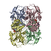
| ||||||||
|---|---|---|---|---|---|---|---|---|---|
| 1 |
| ||||||||
| Unit cell |
|
-Components
| #1: Protein | ( Mass: 36346.551 Da / Num. of mol.: 4 Source method: isolated from a genetically manipulated source Source: (gene. exp.) Candida parapsilosis (fungus) / Strain: CCTCC M203011 / Gene: CPRADH / Plasmid: pET32a / Production host:  #2: Chemical | ChemComp-NAD /   Mass: 663.425 Da / Num. of mol.: 4 / Source method: obtained synthetically / Formula: C21H27N7O14P2 / Comment: NAD*YM Mass: 663.425 Da / Num. of mol.: 4 / Source method: obtained synthetically / Formula: C21H27N7O14P2 / Comment: NAD*YM#3: Chemical | ChemComp-ZN /   Mass: 65.409 Da / Num. of mol.: 8 / Source method: obtained synthetically / Formula: Zn Mass: 65.409 Da / Num. of mol.: 8 / Source method: obtained synthetically / Formula: Zn#4: Water | ChemComp-HOH / |  Mass: 18.015 Da / Num. of mol.: 1076 / Source method: isolated from a natural source / Formula: H2O Mass: 18.015 Da / Num. of mol.: 1076 / Source method: isolated from a natural source / Formula: H2O |
|---|
-Experimental details
-Experiment
| Experiment | Method: X-RAY DIFFRACTION / Number of used crystals: 1 |
|---|
- Sample preparation
| Crystal | Density Matthews: 2.3 Å3/Da / Density % sol: 46.6 % |
|---|---|
| Crystal grow | Temperature: 295 K / Method: vapor diffusion, sitting drop / pH: 7 Details: 0.1M sodium malonate, 12%(w/v) PEG3350, 2%(w/v) PEG10000, pH 7.0, VAPOR DIFFUSION, SITTING DROP, temperature 295K |
-Data collection
| Diffraction | Mean temperature: 100 K |
|---|---|
| Diffraction source | Source: SYNCHROTRON / Site: NSRRC  / Beamline: BL13B1 / Wavelength: 1 Å / Beamline: BL13B1 / Wavelength: 1 Å |
| Detector | Type: ADSC QUANTUM 315 / Detector: CCD / Date: May 5, 2012 |
| Radiation | Monochromator: SI 111 CHANNEL / Protocol: SINGLE WAVELENGTH / Monochromatic (M) / Laue (L): M / Scattering type: x-ray |
| Radiation wavelength | Wavelength: 1 Å / Relative weight: 1 |
| Reflection | Resolution: 2.164→25 Å / Num. all: 72734 / Num. obs: 70570 / % possible obs: 99.9 % / Observed criterion σ(F): 0 / Observed criterion σ(I): 3 / Redundancy: 4.8 % / Rmerge(I) obs: 0.092 / Net I/σ(I): 20.7 |
| Reflection shell | Resolution: 2.164→2.23 Å / Redundancy: 5 % / Rmerge(I) obs: 0.38 / Mean I/σ(I) obs: 5.3 / Num. unique all: 6948 / % possible all: 99.8 |
- Processing
| Software |
| ||||||||||||||||||||
|---|---|---|---|---|---|---|---|---|---|---|---|---|---|---|---|---|---|---|---|---|---|
| Refinement | Method to determine structure: SAD Starting model: PDB ENTRY 2XAA Resolution: 2.164→25 Å / σ(F): 0 / Stereochemistry target values: Engh & Huber
| ||||||||||||||||||||
| Refine analyze |
| ||||||||||||||||||||
| Refinement step | Cycle: LAST / Resolution: 2.164→25 Å
| ||||||||||||||||||||
| Refine LS restraints |
| ||||||||||||||||||||
| LS refinement shell | Resolution: 2.164→2.23 Å
|
