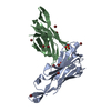
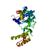
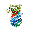
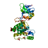
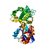
Entry Database : PDB / ID : 4w6wTitle Co-complex structure of the lectin domain of F18 fimbrial adhesin FedF with inhibitory nanobody NbFedF6 F18 fimbrial adhesin AC NbFedF6 Keywords / / / Function / homology / / / / / / Biological species Escherichia coli (E. coli)Lama glama (llama)Method / / / Resolution : 2.51 Å Authors Moonens, K. / De Kerpel, M. / Annelies, C. / Cox, E. / Pardon, E. / Remaut, H. / De Greve, H. Funding support Organization Grant number Country FWO G030411N FWO-Hercules UABR/09/005
Journal : Plos One / Year : 2014Title : Nanobody Mediated Inhibition of Attachment of F18 Fimbriae Expressing Escherichia coli.Authors : Moonens, K. / De Kerpel, M. / Coddens, A. / Cox, E. / Pardon, E. / Remaut, H. / De Greve, H. History Deposition Aug 21, 2014 Deposition site / Processing site Revision 1.0 Dec 17, 2014 Provider / Type Revision 1.1 Dec 24, 2014 Group Revision 1.2 Feb 7, 2018 Group / Category Item _entity_src_gen.gene_src_common_name / _entity_src_gen.pdbx_gene_src_ncbi_taxonomy_id ... _entity_src_gen.gene_src_common_name / _entity_src_gen.pdbx_gene_src_ncbi_taxonomy_id / _entity_src_gen.pdbx_gene_src_scientific_name / _entity_src_gen.pdbx_host_org_ncbi_taxonomy_id / _entity_src_gen.pdbx_host_org_scientific_name / _entity_src_gen.pdbx_host_org_strain / _entity_src_gen.pdbx_host_org_variant / _entity_src_gen.pdbx_host_org_vector_type Revision 1.3 Jan 10, 2024 Group / Database references / Refinement descriptionCategory chem_comp_atom / chem_comp_bond ... chem_comp_atom / chem_comp_bond / database_2 / pdbx_initial_refinement_model Item / _database_2.pdbx_database_accessionRevision 1.4 Nov 6, 2024 Group / Category / pdbx_modification_feature
Show all Show less
 Movie
Movie Controller

 Yorodumi
 Open data
 Basic information
 Components
 Keywords
 Function and homology information

 X-RAY DIFFRACTION /
  Authors
 Belgium, 2items
  Citation
 Structure visualization
 Downloads & links
 Other downloads
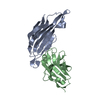
 PDBj
PDBj

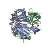
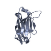
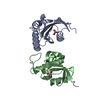
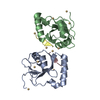
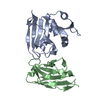
 Assembly
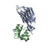
 Mass: 18.015 Da / Num. of mol.: 57 / Source method: isolated from a natural source / Formula: H2O
 Sample preparation
 / Beamline: I04 / Wavelength: 0.98 Å
 Processing