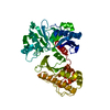
#1: Journal: Microb.Pathog. / Year: 1996 Title: Characterization of F18 Fimbrial Genes Fede and Fedf Involved in Adhesion and Length of Enterotoxemic Escherichia Coli Strain 107/86. Authors: Imberechts, H. / Wild, P. / Charlier, G. / De Greve, H. / Lintermans, P. / Pohl, P.
History
Deposition
Jul 31, 2012
Deposition site: PDBE / Processing site: PDBE
Revision 1.0
Aug 15, 2012
Provider: repository / Type: Initial release
Revision 1.1
Oct 3, 2012
Group: Database references / Source and taxonomy
Revision 1.2
Jul 17, 2019
Group: Data collection / Category: diffrn_source / Item: _diffrn_source.pdbx_synchrotron_site
Protocol: SINGLE WAVELENGTH / Monochromatic (M) / Laue (L): M / Scattering type: x-ray
Radiation wavelength
Wavelength: 1.77 Å / Relative weight: 1
Reflection
Resolution: 1.8→19.81 Å / Num. obs: 24427 / % possible obs: 100 % / Observed criterion σ(I): 2 / Redundancy: 2 % / Rrim(I) all: 0.07 / Net I/σ(I): 28.83
Reflection shell
Highest resolution: 1.8 Å / Mean I/σ(I) obs: 12.94 / Rrim(I) all: 0.185 / % possible all: 100
-
Processing
Software
Name
Version
Classification
SIGMAA
modelbuilding
MLPHARE
modelbuilding
XSCALE
datascaling
SHELXCD
phasing
PHASER
phasing
SIGMAA
phasing
RESOLVE
phasing
BP3
phasing
ABS
phasing
MLPHARE
phasing
REFMAC
5.2.0019
refinement
SHELXE
phasing
Refinement
Method to determine structure: SAD Starting model: NONE Resolution: 1.8→19.81 Å / Cor.coef. Fo:Fc: 0.956 / Cor.coef. Fo:Fc free: 0.928 / SU B: 2.601 / SU ML: 0.082 / Cross valid method: THROUGHOUT / ESU R: 0.129 / ESU R Free: 0.126 / Stereochemistry target values: MAXIMUM LIKELIHOOD / Details: HYDROGENS HAVE BEEN ADDED IN THE RIDING POSITIONS.
Rfactor
Num. reflection
% reflection
Selection details
Rfree
0.22078
1051
4.1 %
RANDOM
Rwork
0.17774
-
-
-
obs
0.17947
24427
99.99 %
-
Solvent computation
Ion probe radii: 0.8 Å / Shrinkage radii: 0.8 Å / VDW probe radii: 1.4 Å / Solvent model: BABINET MODEL WITH MASK
 Movie
Movie Controller

 Yorodumi
 Open data
 Basic information
 Components
 Keywords
 Function and homology information
 X-RAY DIFFRACTION /
  Authors
 Citation
 Structure visualization
 Downloads & links
 Other downloads
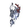


 PDBj
PDBj

 Assembly

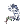
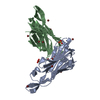
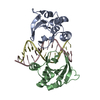
 Mass: 79.904 Da / Num. of mol.: 13 / Source method: obtained synthetically / Formula: Br
 Mass: 96.063 Da / Num. of mol.: 2 / Source method: obtained synthetically / Formula: SO4
 Mass: 18.015 Da / Num. of mol.: 176 / Source method: isolated from a natural source / Formula: H2O
 Sample preparation
 / Beamline: X12 / Wavelength: 1.77
  Processing