 Movie
Movie Controller

[English] 日本語
 Yorodumi
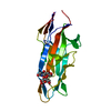
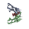
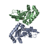
- PDB-4bwo: The FedF adhesin from entrrotoxigenic Escherichia coli is a sulfa... -
+ Open data

- Basic information
| Entry | Database: PDB / ID: 4bwo | ||||||
|---|---|---|---|---|---|---|---|
| Title | The FedF adhesin from entrrotoxigenic Escherichia coli is a sulfate- binding lectin | ||||||
 Components Components | F18 FIMBRIAL ADHESIN AC | ||||||
 Keywords Keywords | CELL ADHESION / GLYCAN ARRAY / ENTEROTOXIGENIC | ||||||
| Function / homology | Jelly Rolls - #1210 / Jelly Rolls / Sandwich / Mainly Beta / BROMIDE ION / F18 fimbrial adhesin AC Function and homology information Function and homology information | ||||||
| Biological species |  | ||||||
| Method |  X-RAY DIFFRACTION / SYNCHROTRON / MAD / Resolution: 1.8 Å X-RAY DIFFRACTION / SYNCHROTRON / MAD / Resolution: 1.8 Å | ||||||
 Authors Authors | Lonardi, E. / Moonens, K. / Buts, L. / de Boer, A.R. / Olsson, J.D.M. / Weiss, M.S. / Fabre, E. / Guerardel, Y. / Deelder, A.M. / Oscarson, S. ...Lonardi, E. / Moonens, K. / Buts, L. / de Boer, A.R. / Olsson, J.D.M. / Weiss, M.S. / Fabre, E. / Guerardel, Y. / Deelder, A.M. / Oscarson, S. / Wuhrer, M. / Bouckaert, J. | ||||||
 Citation Citation | Journal: Mol.Microbiol. / Year: 2012 Title: Structural Insight in Histo-Blood Group Binding by the F18 Fimbrial Adhesin Fedf. Authors: Moonens, K. / Bouckaert, J. / Coddens, A. / Tran, T. / Panjikar, S. / De Kerpel, M. / Cox, E. / Remaut, H. / De Greve, H. #1: Journal: Acta Crystallogr.,Sect.F / Year: 2006 Title: N-Terminal Truncation Enables Crystallization of the Receptor-Binding Domain of the Fedf Bacterial Adhesin. Authors: De Kerpel, M. / Van Molle, I. / Brys, L. / Wyns, L. / De Greve, H. / Bouckaert, J. | ||||||
| History |
| ||||||
| Remark 0 | THIS ENTRY 4BWO REFLECTS AN ALTERNATIVE MODELING OF THE ORIGINAL STRUCTURAL DATA (R4B4PSF) ...THIS ENTRY 4BWO REFLECTS AN ALTERNATIVE MODELING OF THE ORIGINAL STRUCTURAL DATA (R4B4PSF) DETERMINED BY AUTHORS OF THE PDB ENTRY 4B4P: K.MOONENS,J.BOUCKAERT,A.CODDENS,T.TRAN,S.PANJIKAR,M.DE KERPEL,E.COX, H.REMAUT,H.DE GREVE |
- Structure visualization
| Structure viewer | Molecule: MolmilJmol/JSmol |
|---|
- Downloads & links
-Download
| PDBx/mmCIF format | 4bwo.cif.gz | 78 KB | Display | PDBx/mmCIF format |
|---|---|---|---|---|
| PDB format | pdb4bwo.ent.gz | 59 KB | Display | PDB format |
| PDBx/mmJSON format | 4bwo.json.gz | Tree view | PDBx/mmJSON format | |
| Others |  Other downloads Other downloads |
-Validation report
| Arichive directory | https://data.pdbj.org/pub/pdb/validation_reports/bw/4bwoftp://data.pdbj.org/pub/pdb/validation_reports/bw/4bwo | HTTPS FTP |
|---|
-Related structure data


-Links
 PDBj
PDBj

- Assembly
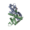
| Deposited unit | 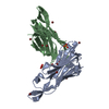
| ||||||||
|---|---|---|---|---|---|---|---|---|---|
| 1 |
| ||||||||
| Unit cell |
|
-Components
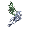
| #1: Protein | Mass: 16311.161 Da / Num. of mol.: 2 / Fragment: LECTIN DOMAIN, RESIDUES 35-185 Source method: isolated from a genetically manipulated source Source: (gene. exp.) #2: Chemical | ChemComp-BR /   Mass: 79.904 Da / Num. of mol.: 13 / Source method: obtained synthetically / Formula: Br Mass: 79.904 Da / Num. of mol.: 13 / Source method: obtained synthetically / Formula: Br#3: Chemical |   Mass: 96.063 Da / Num. of mol.: 3 / Source method: obtained synthetically / Formula: SO4 Mass: 96.063 Da / Num. of mol.: 3 / Source method: obtained synthetically / Formula: SO4#4: Water | ChemComp-HOH / |  Mass: 18.015 Da / Num. of mol.: 286 / Source method: isolated from a natural source / Formula: H2O Mass: 18.015 Da / Num. of mol.: 286 / Source method: isolated from a natural source / Formula: H2OHas protein modification | Y | Nonpolymer details | SULFATE ION (SO4): SULPHATE IONS BOUND OUTSIDE THE SUGAR BINDING SITE BROMIDE ION (BR): BROMINE TO ...SULFATE ION (SO4): SULPHATE IONS BOUND OUTSIDE THE SUGAR BINDING SITE BROMIDE ION (BR): BROMINE TO DETERMINE STRUCTURE USING MAD METHOD | Sequence details | FRAGMENT ENCOMPASSE | |
|---|
-Experimental details
-Experiment
| Experiment | Method: X-RAY DIFFRACTION |
|---|
- Sample preparation
| Crystal | Density Matthews: 1.97 Å3/Da / Density % sol: 37.62 % / Description: AUTHOR USED THE SF DATA FROM ENTRY 4B4P. |
|---|---|
| Crystal grow | Details: 0.17 M AMMONIUM SULFATE, 25.5% (W/V) PEG 4000 AND 15% (V/V) GLYCEROL |
-Data collection
| Diffraction source | Source: SYNCHROTRON / Wavelength: 1 |
|---|---|
| Radiation | Protocol: MAD / Monochromatic (M) / Laue (L): M / Scattering type: x-ray |
| Radiation wavelength | Wavelength: 1 Å / Relative weight: 1 |
| Reflection | Biso Wilson estimate: 18.14 Å2 |
- Processing
| Software |
| |||||||||||||||||||||||||||||||||||||||||||||||||||||||||||||||
|---|---|---|---|---|---|---|---|---|---|---|---|---|---|---|---|---|---|---|---|---|---|---|---|---|---|---|---|---|---|---|---|---|---|---|---|---|---|---|---|---|---|---|---|---|---|---|---|---|---|---|---|---|---|---|---|---|---|---|---|---|---|---|---|---|
| Refinement | Method to determine structure: MAD Starting model: NONE Resolution: 1.8→19.108 Å / SU ML: 0.17 / σ(F): 1.36 / Phase error: 19.1 / Stereochemistry target values: ML
| |||||||||||||||||||||||||||||||||||||||||||||||||||||||||||||||
| Solvent computation | Shrinkage radii: 0.9 Å / VDW probe radii: 1.11 Å / Solvent model: FLAT BULK SOLVENT MODEL / Bsol: 1 Å2 / ksol: 1 e/Å3 | |||||||||||||||||||||||||||||||||||||||||||||||||||||||||||||||
| Displacement parameters | Biso mean: 22 Å2 | |||||||||||||||||||||||||||||||||||||||||||||||||||||||||||||||
| Refinement step | Cycle: LAST / Resolution: 1.8→19.108 Å
| |||||||||||||||||||||||||||||||||||||||||||||||||||||||||||||||
| Refine LS restraints |
| |||||||||||||||||||||||||||||||||||||||||||||||||||||||||||||||
| LS refinement shell |
|
