| Entry | Database: PDB / ID: 4o9i
|
|---|
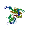
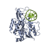
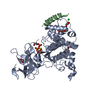
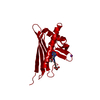
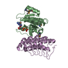
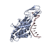
| Title | Structure of CHD4 double chromodomains depicts cooperative folding for DNA binding |
|---|
 Components Components | Chromodomain-helicase-DNA-binding protein 4 |
|---|
 Keywords Keywords | HYDROLASE / CHD4 double chromodomains |
|---|
| Function / homology |  Function and homology information Function and homology information
cerebellar granule cell to Purkinje cell synapse / terminal button organization / NuRD complex / regulation of cell fate specification / NGF-stimulated transcription / regulation of stem cell differentiation / ATP-dependent chromatin remodeler activity / regulation of synapse assembly / RNA Polymerase I Transcription Initiation / Transcriptional regulation of brown and beige adipocyte differentiation by EBF2 ...cerebellar granule cell to Purkinje cell synapse / terminal button organization / NuRD complex / regulation of cell fate specification / NGF-stimulated transcription / regulation of stem cell differentiation / ATP-dependent chromatin remodeler activity / regulation of synapse assembly / RNA Polymerase I Transcription Initiation / Transcriptional regulation of brown and beige adipocyte differentiation by EBF2 / Regulation of TP53 Activity through Acetylation / site of DNA damage / Regulation of PTEN gene transcription / transcription coregulator binding / ERCC6 (CSB) and EHMT2 (G9a) positively regulate rRNA expression / Regulation of endogenous retroelements by KRAB-ZFP proteins / HDACs deacetylate histones / Regulation of endogenous retroelements by Piwi-interacting RNAs (piRNAs) / double-strand break repair via homologous recombination / RNA polymerase II transcription regulator complex / Hydrolases; Acting on acid anhydrides; Acting on acid anhydrides to facilitate cellular and subcellular movement / histone deacetylase binding / transcription corepressor activity / histone binding / Potential therapeutics for SARS / RNA polymerase II-specific DNA-binding transcription factor binding / chromosome, telomeric region / chromatin remodeling / negative regulation of gene expression / negative regulation of DNA-templated transcription / chromatin binding / centrosome / positive regulation of DNA-templated transcription / chromatin / negative regulation of transcription by RNA polymerase II / ATP hydrolysis activity / protein-containing complex / DNA binding / zinc ion binding / nucleoplasm / ATP binding / membrane / nucleus / cytoplasmSimilarity search - Function CHD subfamily II, SANT-like domain / Domain of unknown function DUF1087 / CHD, C-terminal 2 / CHD, N-terminal / CHD subfamily II, SANT-like domain / CHD subfamily II, DUF1087 / CHDNT (NUC034) domain / CHDCT2 (NUC038) domain / Domain of Unknown Function (DUF1086) / DUF1087 ...CHD subfamily II, SANT-like domain / Domain of unknown function DUF1087 / CHD, C-terminal 2 / CHD, N-terminal / CHD subfamily II, SANT-like domain / CHD subfamily II, DUF1087 / CHDNT (NUC034) domain / CHDCT2 (NUC038) domain / Domain of Unknown Function (DUF1086) / DUF1087 / Chromo domain signature. / Chromo domain / Chromo (CHRromatin Organisation MOdifier) domain / Chromo and chromo shadow domain profile. / Chromo/chromo shadow domain / Chromatin organization modifier domain / Chromo-like domain superfamily / DNA/RNA helicase, ATP-dependent, DEAH-box type, conserved site / DEAH-box subfamily ATP-dependent helicases signature. / : / SNF2-like, N-terminal domain superfamily / SNF2, N-terminal / SNF2-related domain / Zinc finger, PHD-type, conserved site / PHD-finger / Zinc finger PHD-type signature. / Zinc finger PHD-type profile. / Zinc finger, PHD-finger / Zinc finger, PHD-type / PHD zinc finger / Zinc finger, FYVE/PHD-type / Helicase conserved C-terminal domain / helicase superfamily c-terminal domain / Superfamilies 1 and 2 helicase C-terminal domain profile. / Superfamilies 1 and 2 helicase ATP-binding type-1 domain profile. / DEAD-like helicases superfamily / Helicase, C-terminal / Helicase superfamily 1/2, ATP-binding domain / Zinc finger, RING/FYVE/PHD-type / P-loop containing nucleoside triphosphate hydrolaseSimilarity search - Domain/homology |
|---|
| Biological species |  Homo sapiens (human) Homo sapiens (human) |
|---|
| Method |  X-RAY DIFFRACTION / SYNCHROTRON / SAD / Resolution: 2.6 Å X-RAY DIFFRACTION / SYNCHROTRON / SAD / Resolution: 2.6 Å |
|---|
 Authors Authors | Wiggs, K.R. / Chruszcz, M. / Su, X. / Minor, W. / Khorasanizadeh, S. |
|---|
 Citation Citation | Journal: TO BE PUBLISHED
Title: Structure of CHD4 double chromodomains depicts cooperative folding for DNA binding
Authors: Wiggs, K.R. / Chruszcz, M. / Su, X. / Minor, W. / Khorasanizadeh, S. |
|---|
| History | | Deposition | Jan 2, 2014 | Deposition site: RCSB / Processing site: RCSB |
|---|
| Revision 1.0 | Jul 8, 2015 | Provider: repository / Type: Initial release |
|---|
| Revision 1.1 | Nov 22, 2017 | Group: Refinement description / Category: software
Item: _software.classification / _software.name / _software.version |
|---|
| Revision 1.2 | Apr 13, 2022 | Group: Database references / Derived calculations / Structure summary
Category: audit_author / citation_author ...audit_author / citation_author / database_2 / struct_conn / struct_ref_seq_dif
Item: _audit_author.identifier_ORCID / _citation_author.identifier_ORCID ..._audit_author.identifier_ORCID / _citation_author.identifier_ORCID / _database_2.pdbx_DOI / _database_2.pdbx_database_accession / _struct_conn.pdbx_leaving_atom_flag / _struct_ref_seq_dif.details |
|---|
| Revision 1.3 | Nov 6, 2024 | Group: Data collection / Structure summary
Category: chem_comp_atom / chem_comp_bond ...chem_comp_atom / chem_comp_bond / pdbx_entry_details / pdbx_modification_feature |
|---|
|
|---|
 Movie
Movie Controller

 Yorodumi
 Open data
 Basic information
 Structure visualization
 Downloads & links
 Other downloads
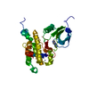

 PDBj
PDBj


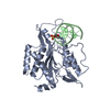
 Assembly

 Mass: 18.015 Da / Num. of mol.: 25 / Source method: isolated from a natural source / Formula: H2O
 Sample preparation
 / Beamline: 22-ID / Wavelength: 0.97907 Å
 Processing