 Movie
Movie Controller

[English] 日本語
 Yorodumi
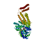
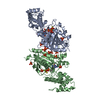
- PDB-3epu: Crystal Structure of STM2138, a novel virulence chaperone in Salm... -
+ Open data

- Basic information
| Entry | Database: PDB / ID: 3epu | ||||||
|---|---|---|---|---|---|---|---|
| Title | Crystal Structure of STM2138, a novel virulence chaperone in Salmonella | ||||||
 Components Components | STM2138 Virulence Chaperone | ||||||
 Keywords Keywords | CHAPERONE / Virulence Chaperone / Salmonella / TypeIII Secretion System / STM2138 / SrcA (SsrB-regulated chaperone A) | ||||||
| Function / homology |  Function and homology information Function and homology information | ||||||
| Biological species |  Salmonella typhimurium (bacteria) Salmonella typhimurium (bacteria) | ||||||
| Method |  X-RAY DIFFRACTION / SYNCHROTRON / MOLECULAR REPLACEMENT / Resolution: 2.5 Å X-RAY DIFFRACTION / SYNCHROTRON / MOLECULAR REPLACEMENT / Resolution: 2.5 Å | ||||||
 Authors Authors | Zhang, K. / Andres, S.N. / Hannemann, M. / Coombes, B. / Junop, M. | ||||||
 Citation Citation | Journal: To be Published Title: Structural analysis and quantitative proteomic interactome of a novel virulence chaperone in Salmonella Authors: Cooper, C. / Zhang, K. / Andres, S.N. / Hannemann, M. / Junop, M.S. / Coombes, B. | ||||||
| History |
|
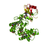
- Structure visualization
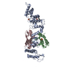
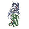
| Structure viewer | Molecule: MolmilJmol/JSmol |
|---|
- Downloads & links
-Download
| PDBx/mmCIF format | 3epu.cif.gz | 125.3 KB | Display | PDBx/mmCIF format |
|---|---|---|---|---|
| PDB format | pdb3epu.ent.gz | 99.3 KB | Display | PDB format |
| PDBx/mmJSON format | 3epu.json.gz | Tree view | PDBx/mmJSON format | |
| Others |  Other downloads Other downloads |
-Validation report
| Arichive directory | https://data.pdbj.org/pub/pdb/validation_reports/ep/3epuftp://data.pdbj.org/pub/pdb/validation_reports/ep/3epu | HTTPS FTP |
|---|
-Related structure data
| Related structure data | 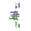 1k3eS S: Starting model for refinement |
|---|---|
| Similar structure data |

-Links
 PDBj
PDBj
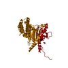
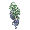
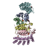
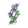
- Assembly
| Deposited unit | 
| ||||||||
|---|---|---|---|---|---|---|---|---|---|
| 1 | 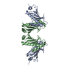
| ||||||||
| 2 |
| ||||||||
| Unit cell |
|
-Components
| #1: Protein | Mass: 16906.959 Da / Num. of mol.: 2 Source method: isolated from a genetically manipulated source Source: (gene. exp.) Salmonella typhimurium (bacteria) / Gene: STM2138 / Plasmid: pET3a / Production host: #2: Water | ChemComp-HOH / |  Mass: 18.015 Da / Num. of mol.: 86 / Source method: isolated from a natural source / Formula: H2O Mass: 18.015 Da / Num. of mol.: 86 / Source method: isolated from a natural source / Formula: H2O |
|---|
-Experimental details
-Experiment
| Experiment | Method: X-RAY DIFFRACTION / Number of used crystals: 1 |
|---|
- Sample preparation
| Crystal | Density Matthews: 2.27 Å3/Da / Density % sol: 45.92 % |
|---|---|
| Crystal grow | Temperature: 298 K / Method: vapor diffusion, hanging drop / pH: 7 Details: 100 mM Bis-Tris propane, 200 mM MgCl2, 35% PEG 3350, 3.95 mM FOS-choline-9, 5% (v/v) Jeffamine M-600, pH 7.0, VAPOR DIFFUSION, HANGING DROP, temperature 298.0K |
-Data collection
| Diffraction | Mean temperature: 100 K |
|---|---|
| Diffraction source | Source: SYNCHROTRON / Site: NSLS  / Beamline: X12C / Wavelength: 1.1 Å / Beamline: X12C / Wavelength: 1.1 Å |
| Detector | Type: ADSC QUANTUM 210 / Detector: CCD / Date: Apr 24, 2008 / Details: toroidal mirror |
| Radiation | Monochromator: Si(111) channel-cut crystal monochromator / Protocol: SINGLE WAVELENGTH / Monochromatic (M) / Laue (L): M / Scattering type: x-ray |
| Radiation wavelength | Wavelength: 1.1 Å / Relative weight: 1 |
| Reflection | Resolution: 2.49→50 Å / Num. all: 10664 / Num. obs: 10664 / % possible obs: 98.5 % / Observed criterion σ(F): 1 / Observed criterion σ(I): 1 / Redundancy: 3.62 % / Biso Wilson estimate: 54.8 Å2 / Rmerge(I) obs: 0.064 / Net I/σ(I): 13.5 |
| Reflection shell | Resolution: 2.49→2.58 Å / Redundancy: 3.58 % / Rmerge(I) obs: 0.316 / Mean I/σ(I) obs: 3.4 / % possible all: 94.6 |
- Processing
| Software |
| ||||||||||||||||||||||||||||||||||||||||||||||||||||||||||||||||||||||||||||||||||||||||||||||||||||||||||||||||||||||||||||||||||||||||||||||||||||||
|---|---|---|---|---|---|---|---|---|---|---|---|---|---|---|---|---|---|---|---|---|---|---|---|---|---|---|---|---|---|---|---|---|---|---|---|---|---|---|---|---|---|---|---|---|---|---|---|---|---|---|---|---|---|---|---|---|---|---|---|---|---|---|---|---|---|---|---|---|---|---|---|---|---|---|---|---|---|---|---|---|---|---|---|---|---|---|---|---|---|---|---|---|---|---|---|---|---|---|---|---|---|---|---|---|---|---|---|---|---|---|---|---|---|---|---|---|---|---|---|---|---|---|---|---|---|---|---|---|---|---|---|---|---|---|---|---|---|---|---|---|---|---|---|---|---|---|---|---|---|---|---|
| Refinement | Method to determine structure: MOLECULAR REPLACEMENT Starting model: PDB entry 1K3E Resolution: 2.5→33.4 Å / Cor.coef. Fo:Fc: 0.938 / Cor.coef. Fo:Fc free: 0.931 / SU B: 16.719 / SU ML: 0.172 / Cross valid method: THROUGHOUT / σ(F): 1 / σ(I): 1 / ESU R Free: 0.303 / Stereochemistry target values: MAXIMUM LIKELIHOOD Details: HYDROGENS HAVE BEEN ADDED IN THE RIDING POSITIONS. THE FIRST 2 TLS GROUPS CORRESPONDS TO THE MAIN CHAIN ATOMS AND THE TLS-3,4 CORRESPONDS TO THE SIDE-CHAINS OF THE SAME RESIDUES
| ||||||||||||||||||||||||||||||||||||||||||||||||||||||||||||||||||||||||||||||||||||||||||||||||||||||||||||||||||||||||||||||||||||||||||||||||||||||
| Solvent computation | Ion probe radii: 0.8 Å / Shrinkage radii: 0.8 Å / VDW probe radii: 1.4 Å / Solvent model: BABINET MODEL WITH MASK | ||||||||||||||||||||||||||||||||||||||||||||||||||||||||||||||||||||||||||||||||||||||||||||||||||||||||||||||||||||||||||||||||||||||||||||||||||||||
| Displacement parameters | Biso mean: 63.824 Å2
| ||||||||||||||||||||||||||||||||||||||||||||||||||||||||||||||||||||||||||||||||||||||||||||||||||||||||||||||||||||||||||||||||||||||||||||||||||||||
| Refinement step | Cycle: LAST / Resolution: 2.5→33.4 Å
| ||||||||||||||||||||||||||||||||||||||||||||||||||||||||||||||||||||||||||||||||||||||||||||||||||||||||||||||||||||||||||||||||||||||||||||||||||||||
| Refine LS restraints |
| ||||||||||||||||||||||||||||||||||||||||||||||||||||||||||||||||||||||||||||||||||||||||||||||||||||||||||||||||||||||||||||||||||||||||||||||||||||||
| LS refinement shell | Resolution: 2.5→2.565 Å / Total num. of bins used: 20
| ||||||||||||||||||||||||||||||||||||||||||||||||||||||||||||||||||||||||||||||||||||||||||||||||||||||||||||||||||||||||||||||||||||||||||||||||||||||
| Refinement TLS params. | Method: refined / Refine-ID: X-RAY DIFFRACTION
| ||||||||||||||||||||||||||||||||||||||||||||||||||||||||||||||||||||||||||||||||||||||||||||||||||||||||||||||||||||||||||||||||||||||||||||||||||||||
| Refinement TLS group |
|
