 Movie
Movie Controller


+ Open data

- Basic information
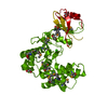
| Entry | Database: PDB / ID: 1azt | ||||||
|---|---|---|---|---|---|---|---|
| Title | GS-ALPHA COMPLEXED WITH GTP-GAMMA-S | ||||||
 Components Components | GS-ALPHA | ||||||
 Keywords Keywords | HYDROLASE / SIGNAL TRANSDUCING PROTEIN / GTP-BINDING PROTEIN | ||||||
| Function / homology |  Function and homology information Function and homology informationsensory perception of chemical stimulus / mu-type opioid receptor binding / corticotropin-releasing hormone receptor 1 binding / beta-2 adrenergic receptor binding / D1 dopamine receptor binding / adenylate cyclase-activating adrenergic receptor signaling pathway / insulin-like growth factor receptor binding / ionotropic glutamate receptor binding / adenylate cyclase activator activity / G-protein beta/gamma-subunit complex binding ...sensory perception of chemical stimulus / mu-type opioid receptor binding / corticotropin-releasing hormone receptor 1 binding / beta-2 adrenergic receptor binding / D1 dopamine receptor binding / adenylate cyclase-activating adrenergic receptor signaling pathway / insulin-like growth factor receptor binding / ionotropic glutamate receptor binding / adenylate cyclase activator activity / G-protein beta/gamma-subunit complex binding / adenylate cyclase-activating dopamine receptor signaling pathway / heterotrimeric G-protein complex / adenylate cyclase-activating G protein-coupled receptor signaling pathway / Hydrolases; Acting on acid anhydrides; Acting on GTP to facilitate cellular and subcellular movement / GTPase activity / GTP binding / metal ion binding / cytoplasm Similarity search - Function | ||||||
| Biological species |  | ||||||
| Method |  X-RAY DIFFRACTION / SYNCHROTRON / MOLECULAR REPLACEMENT / Resolution: 2.3 Å X-RAY DIFFRACTION / SYNCHROTRON / MOLECULAR REPLACEMENT / Resolution: 2.3 Å | ||||||
 Authors Authors | Tesmer, J.J.G. / Sprang, S.R. | ||||||
 Citation Citation | Journal: Science / Year: 1997 Title: Crystal structure of the adenylyl cyclase activator Gsalpha Authors: Sunahara, R.K. / Tesmer, J.J. / Gilman, A.G. / Sprang, S.R. | ||||||
| History |
|
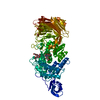
- Structure visualization
| Structure viewer | Molecule: MolmilJmol/JSmol |
|---|
- Downloads & links
-Download
| PDBx/mmCIF format | 1azt.cif.gz | 155.3 KB | Display | PDBx/mmCIF format |
|---|---|---|---|---|
| PDB format | pdb1azt.ent.gz | 121.9 KB | Display | PDB format |
| PDBx/mmJSON format | 1azt.json.gz | Tree view | PDBx/mmJSON format | |
| Others |  Other downloads Other downloads |
-Validation report
| Arichive directory | https://data.pdbj.org/pub/pdb/validation_reports/az/1aztftp://data.pdbj.org/pub/pdb/validation_reports/az/1azt | HTTPS FTP |
|---|
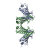
-Related structure data
| Related structure data |  1giaS S: Starting model for refinement |
|---|---|
| Similar structure data |


-Links
 PDBj
PDBj


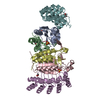
- Assembly
| Deposited unit | 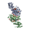
| ||||||||
|---|---|---|---|---|---|---|---|---|---|
| 1 |
| ||||||||
| Unit cell |
| ||||||||
| Noncrystallographic symmetry (NCS) | NCS oper: (Code: given Matrix: (0.9999, -0.0113, -0.0105), Vector: |
-Components
| #1: Protein | Mass: 46712.500 Da / Num. of mol.: 2 Mutation: C-TERMINAL HEXAHISTIDINE TAG, NOT PALMITOYLATED AT AMINO TERMINUS Source method: isolated from a genetically manipulated source Source: (gene. exp.)  #2: Chemical |   Mass: 24.305 Da / Num. of mol.: 2 / Source method: obtained synthetically / Formula: Mg Mass: 24.305 Da / Num. of mol.: 2 / Source method: obtained synthetically / Formula: Mg#3: Chemical | ChemComp-PO4 /   Mass: 94.971 Da / Num. of mol.: 16 / Source method: obtained synthetically / Formula: PO4 Mass: 94.971 Da / Num. of mol.: 16 / Source method: obtained synthetically / Formula: PO4#4: Chemical |   Mass: 539.246 Da / Num. of mol.: 2 / Source method: obtained synthetically / Formula: C10H16N5O13P3S Mass: 539.246 Da / Num. of mol.: 2 / Source method: obtained synthetically / Formula: C10H16N5O13P3S#5: Water | ChemComp-HOH / |  Mass: 18.015 Da / Num. of mol.: 57 / Source method: isolated from a natural source / Formula: H2O Mass: 18.015 Da / Num. of mol.: 57 / Source method: isolated from a natural source / Formula: H2OSequence details | DATABASE ENTRY IS FOR THE LONG SPLICE VARIANT. RESIDUES 71-84 ARE SPLICED OUT IN THE SHORT SPLICE ...DATABASE ENTRY IS FOR THE LONG SPLICE VARIANT. RESIDUES 71-84 ARE SPLICED OUT IN THE SHORT SPLICE VARIANT USED IN THIS EXPERIMENT | |
|---|
-Experimental details
-Experiment
| Experiment | Method: X-RAY DIFFRACTION / Number of used crystals: 2 |
|---|
- Sample preparation
| Crystal | Density Matthews: 3.1 Å3/Da / Density % sol: 60 % | ||||||||||||||||||||||||||||||||||||||||||||||||||||||||
|---|---|---|---|---|---|---|---|---|---|---|---|---|---|---|---|---|---|---|---|---|---|---|---|---|---|---|---|---|---|---|---|---|---|---|---|---|---|---|---|---|---|---|---|---|---|---|---|---|---|---|---|---|---|---|---|---|---|
| Crystal grow | Method: vapor diffusion, hanging drop / pH: 4.5 Details: CRYSTALLIZED IN HANGING DROPS CONTAINING PROTEIN MIXED 1:1 WITH WELL SOLUTION CONSISTING OF 90-100% SATURATED KH2PO4 OR NAH2PO4 (UNBUFFERED)., pH 4.5, vapor diffusion - hanging drop | ||||||||||||||||||||||||||||||||||||||||||||||||||||||||
| Crystal | *PLUS | ||||||||||||||||||||||||||||||||||||||||||||||||||||||||
| Crystal grow | *PLUS Temperature: 20 ℃ / pH: 8 / Method: vapor diffusion, hanging drop | ||||||||||||||||||||||||||||||||||||||||||||||||||||||||
| Components of the solutions | *PLUS
|
-Data collection
| Diffraction | Mean temperature: 100 K |
|---|---|
| Diffraction source | Source: SYNCHROTRON / Site: CHESS  / Beamline: A1 / Wavelength: 0.908 / Beamline: A1 / Wavelength: 0.908 |
| Detector | Type: ADSC QUANTUM / Detector: CCD / Date: Jun 1, 1997 |
| Radiation | Monochromatic (M) / Laue (L): M / Scattering type: x-ray |
| Radiation wavelength | Wavelength: 0.908 Å / Relative weight: 1 |
| Reflection | Highest resolution: 2.5 Å / Num. obs: 37636 / % possible obs: 94 % / Observed criterion σ(I): -2 / Redundancy: 4.2 % / Biso Wilson estimate: 30.6 Å2 / Rsym value: 0.138 / Net I/σ(I): 8.3 |
| Reflection shell | Resolution: 2.5→2.54 Å / Redundancy: 2.7 % / Mean I/σ(I) obs: 2.5 / Rsym value: 0.417 / % possible all: 95.1 |
| Reflection | *PLUS Rmerge(I) obs: 0.138 |
- Processing
| Software |
| ||||||||||||||||||||||||||||||||||||||||||||||||||||||||||||||||||||||||||||||||
|---|---|---|---|---|---|---|---|---|---|---|---|---|---|---|---|---|---|---|---|---|---|---|---|---|---|---|---|---|---|---|---|---|---|---|---|---|---|---|---|---|---|---|---|---|---|---|---|---|---|---|---|---|---|---|---|---|---|---|---|---|---|---|---|---|---|---|---|---|---|---|---|---|---|---|---|---|---|---|---|---|---|
| Refinement | Method to determine structure: MOLECULAR REPLACEMENT Starting model: PDB ENTRY 1GIA Resolution: 2.3→15 Å / Rfactor Rfree error: 0.005 / Isotropic thermal model: RESTRAINED / Cross valid method: THROUGHOUT / Details: BULK SOLVENT CORRECTION WAS USED.
| ||||||||||||||||||||||||||||||||||||||||||||||||||||||||||||||||||||||||||||||||
| Displacement parameters | Biso mean: 52.2 Å2
| ||||||||||||||||||||||||||||||||||||||||||||||||||||||||||||||||||||||||||||||||
| Refine analyze | Luzzati d res low obs: 15 Å / Luzzati sigma a obs: 0.31 Å | ||||||||||||||||||||||||||||||||||||||||||||||||||||||||||||||||||||||||||||||||
| Refinement step | Cycle: LAST / Resolution: 2.3→15 Å
| ||||||||||||||||||||||||||||||||||||||||||||||||||||||||||||||||||||||||||||||||
| Refine LS restraints |
| ||||||||||||||||||||||||||||||||||||||||||||||||||||||||||||||||||||||||||||||||
| LS refinement shell | Resolution: 2.3→2.4 Å / Rfactor Rfree error: 0.022 / Total num. of bins used: 8
| ||||||||||||||||||||||||||||||||||||||||||||||||||||||||||||||||||||||||||||||||
| Xplor file |
| ||||||||||||||||||||||||||||||||||||||||||||||||||||||||||||||||||||||||||||||||
| Software | *PLUS Name: X-PLOR / Version: 3.851 / Classification: refinement | ||||||||||||||||||||||||||||||||||||||||||||||||||||||||||||||||||||||||||||||||
| Refine LS restraints | *PLUS
|
