 Movie
Movie Controller

[English] 日本語
 Yorodumi
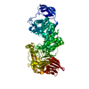
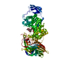
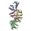
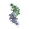
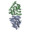
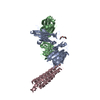
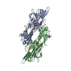
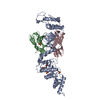
- PDB-1i8q: CRYSTAL STRUCTURE OF STREPTOCOCCUS AGALACTIAE HYALURONATE LYASE C... -
+ Open data

- Basic information
| Entry | Database: PDB / ID: 1i8q | |||||||||
|---|---|---|---|---|---|---|---|---|---|---|
| Title | CRYSTAL STRUCTURE OF STREPTOCOCCUS AGALACTIAE HYALURONATE LYASE COMPLEXED WITH ENZYME PRODUCT, UNSATURATED DISACCHARIDE HYALURONAN | |||||||||
 Components Components | HYALURONATE LYASE | |||||||||
 Keywords Keywords | LYASE / beta-alpha-beta | |||||||||
| Function / homology |  Function and homology information Function and homology informationhyaluronate lyase / hyaluronate lyase activity / carbohydrate binding / carbohydrate metabolic process / extracellular region Similarity search - Function | |||||||||
| Biological species |  Streptococcus agalactiae (bacteria) Streptococcus agalactiae (bacteria) | |||||||||
| Method |  X-RAY DIFFRACTION / SYNCHROTRON / MOLECULAR REPLACEMENT / Resolution: 2.2 Å X-RAY DIFFRACTION / SYNCHROTRON / MOLECULAR REPLACEMENT / Resolution: 2.2 Å | |||||||||
 Authors Authors | Li, S. / Jedrzejas, M.J. | |||||||||
 Citation Citation | Journal: J.Biol.Chem. / Year: 2001 Title: Hyaluronan binding and degradation by Streptococcus agalactiae hyaluronate lyase. Authors: Li, S. / Jedrzejas, M.J. | |||||||||
| History |
|
- Structure visualization
| Structure viewer | Molecule: MolmilJmol/JSmol |
|---|
- Downloads & links
-Download
| PDBx/mmCIF format | 1i8q.cif.gz | 180.4 KB | Display | PDBx/mmCIF format |
|---|---|---|---|---|
| PDB format | pdb1i8q.ent.gz | 140.2 KB | Display | PDB format |
| PDBx/mmJSON format | 1i8q.json.gz | Tree view | PDBx/mmJSON format | |
| Others |  Other downloads Other downloads |
-Validation report
| Arichive directory | https://data.pdbj.org/pub/pdb/validation_reports/i8/1i8qftp://data.pdbj.org/pub/pdb/validation_reports/i8/1i8q | HTTPS FTP |
|---|
-Related structure data


-Links
 PDBj
PDBj


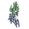
- Assembly
| Deposited unit | 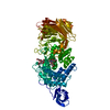
| ||||||||
|---|---|---|---|---|---|---|---|---|---|
| 1 |
| ||||||||
| Unit cell |
|
-Components
| #1: Protein | Mass: 92510.344 Da / Num. of mol.: 1 / Fragment: ENZYMATICALLY ACTIVE FRAGMENT, RESIDUES 171-984 Source method: isolated from a genetically manipulated source Source: (gene. exp.) Streptococcus agalactiae (bacteria) / Gene: HYL / Production host: | ||
|---|---|---|---|
| #2: Polysaccharide | Source method: isolated from a genetically manipulated source #3: Water | ChemComp-HOH / |  Mass: 18.015 Da / Num. of mol.: 229 / Source method: isolated from a natural source / Formula: H2O Mass: 18.015 Da / Num. of mol.: 229 / Source method: isolated from a natural source / Formula: H2O |
-Experimental details
-Experiment
| Experiment | Method: X-RAY DIFFRACTION / Number of used crystals: 1 |
|---|
- Sample preparation
| Crystal | Density Matthews: 2.54 Å3/Da / Density % sol: 51.49 % | ||||||||||||||||||||||||||||||||||||||||||
|---|---|---|---|---|---|---|---|---|---|---|---|---|---|---|---|---|---|---|---|---|---|---|---|---|---|---|---|---|---|---|---|---|---|---|---|---|---|---|---|---|---|---|---|
| Crystal grow | Temperature: 298 K / Method: vapor diffusion, hanging drop / pH: 6 Details: PEG 5500 MME, KSCN, CACODYLATE, pH 6.0, VAPOR DIFFUSION, HANGING DROP, temperature 298K | ||||||||||||||||||||||||||||||||||||||||||
| Crystal grow | *PLUS pH: 8 Details: Jedrzejas, M.J., (2000) Acta Crystallogr., Sect.D, 56, 460. | ||||||||||||||||||||||||||||||||||||||||||
| Components of the solutions | *PLUS
|
-Data collection
| Diffraction | Mean temperature: 100 K |
|---|---|
| Diffraction source | Source: SYNCHROTRON / Site: APS  / Beamline: 19-ID / Wavelength: 0.98 Å / Beamline: 19-ID / Wavelength: 0.98 Å |
| Detector | Detector: CCD / Date: Sep 25, 2000 |
| Radiation | Protocol: SINGLE WAVELENGTH / Monochromatic (M) / Laue (L): M / Scattering type: x-ray |
| Radiation wavelength | Wavelength: 0.98 Å / Relative weight: 1 |
| Reflection | Resolution: 2.2→50 Å / Num. obs: 47142 / % possible obs: 98.1 % / Observed criterion σ(I): 5 / Redundancy: 14.1 % / Biso Wilson estimate: 36.296 Å2 / Rmerge(I) obs: 0.123 / Net I/σ(I): 10.5 |
| Reflection | *PLUS Num. obs: 45510 / Num. measured all: 295197 |
| Reflection shell | *PLUS % possible obs: 91.5 % / Rmerge(I) obs: 0.216 |
- Processing
| Software |
| ||||||||||||||||||||||||||||||
|---|---|---|---|---|---|---|---|---|---|---|---|---|---|---|---|---|---|---|---|---|---|---|---|---|---|---|---|---|---|---|---|
| Refinement | Method to determine structure: MOLECULAR REPLACEMENT Starting model: native structure of streptococcus agalactiae hyaluronate lyase Resolution: 2.2→20 Å / Cross valid method: THROUGHOUT / σ(F): 0.001 / σ(I): 0.001 / Stereochemistry target values: Engh & Huber
| ||||||||||||||||||||||||||||||
| Refinement step | Cycle: LAST / Resolution: 2.2→20 Å
| ||||||||||||||||||||||||||||||
| Software | *PLUS Name: X-PLOR / Version: 3.851 / Classification: refinement | ||||||||||||||||||||||||||||||
| Refinement | *PLUS Highest resolution: 2.2 Å / Lowest resolution: 20 Å / Rfactor obs: 0.182 | ||||||||||||||||||||||||||||||
| Solvent computation | *PLUS | ||||||||||||||||||||||||||||||
| Displacement parameters | *PLUS | ||||||||||||||||||||||||||||||
| Refine LS restraints | *PLUS
|
