 Movie
Movie Controller

[English] 日本語
 Yorodumi
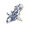
- PDB-1eri: X-RAY STRUCTURE OF THE DNA-ECO RI ENDONUCLEASE-DNA RECOGNITION CO... -
+ Open data

- Basic information
| Entry | Database: PDB / ID: 1eri | |||||||||
|---|---|---|---|---|---|---|---|---|---|---|
| Title | X-RAY STRUCTURE OF THE DNA-ECO RI ENDONUCLEASE-DNA RECOGNITION COMPLEX: THE RECOGNITION NETWORK AND THE INTEGRATION OF RECOGNITION AND CLEAVAGE | |||||||||
 Components Components |
| |||||||||
 Keywords Keywords | HYDROLASE/DNA / PROTEIN-DNA COMPLEX / DOUBLE HELIX / HYDROLASE-DNA COMPLEX | |||||||||
| Function / homology |  Function and homology information Function and homology informationtype II site-specific deoxyribonuclease / type II site-specific deoxyribonuclease activity / DNA restriction-modification system / magnesium ion binding / DNA binding Similarity search - Function | |||||||||
| Biological species |  | |||||||||
| Method |  X-RAY DIFFRACTION / Resolution: 2.5 Å X-RAY DIFFRACTION / Resolution: 2.5 Å | |||||||||
 Authors Authors | Kim, Y. / Grable, J.C. / Love, R. / Greene, P.J. / Rosenberg, J.M. | |||||||||
 Citation Citation | Journal: Science / Year: 1990 Title: Refinement of Eco RI endonuclease crystal structure: a revised protein chain tracing. Authors: Kim, Y.C. / Grable, J.C. / Love, R. / Greene, P.J. / Rosenberg, J.M. #1: Journal: To be PublishedTitle: Preliminary Characterization of EcoRI-DNA Co-Crystals: Incomplete Factorial Design of Oligonucleotide Sequences Authors: Wilkosz, P.A. / Chandrasekhar, K. / Rosenberg, J.M. #2: Journal: To be PublishedTitle: Molecular Dynamics Simulations Suggest that the EcoRI Kink is an Example of Molecular Strain Authors: Kumar, S. / Duan, Y. / Kollman, P.A. / Rosenberg, J.M. #3: Journal: Structural Biology: The State of the Art; Proceedings of the Eighth Conversation, State University of New York, AlbanyYear: 1994 Title: Studies on the Canonical DNA-EcoRI Endonuclease Complex and the EcoRI Kink Authors: Kim, Y. / Choi, J. / Grable, J.C. / Greene, P. / Hager, P. / Rosenberg, J.M. #4: Journal: Curr.Opin.Struct.Biol. / Year: 1991Title: Structure and Function of Restriction Endonucleases Authors: Rosenberg, J.M. | |||||||||
| History |
|
- Structure visualization
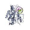
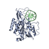
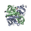
| Structure viewer | Molecule: MolmilJmol/JSmol |
|---|
- Downloads & links
-Download
| PDBx/mmCIF format | 1eri.cif.gz | 74.7 KB | Display | PDBx/mmCIF format |
|---|---|---|---|---|
| PDB format | pdb1eri.ent.gz | 52.9 KB | Display | PDB format |
| PDBx/mmJSON format | 1eri.json.gz | Tree view | PDBx/mmJSON format | |
| Others |  Other downloads Other downloads |
-Validation report
| Arichive directory | https://data.pdbj.org/pub/pdb/validation_reports/er/1eriftp://data.pdbj.org/pub/pdb/validation_reports/er/1eri | HTTPS FTP |
|---|
-Related structure data
| Similar structure data |
|---|


-Links
 PDBj
PDBj


- Assembly
| Deposited unit | 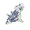
| ||||||||
|---|---|---|---|---|---|---|---|---|---|
| 1 | 
| ||||||||
| Unit cell |
|
-Components
| #1: DNA chain | Mass: 3967.585 Da / Num. of mol.: 1 / Source method: obtained synthetically |
|---|---|
| #2: Protein | Mass: 30970.184 Da / Num. of mol.: 1 / Source method: isolated from a natural source / Source: (natural) |
| #3: Water | ChemComp-HOH /  Mass: 18.015 Da / Num. of mol.: 61 / Source method: isolated from a natural source / Formula: H2O Mass: 18.015 Da / Num. of mol.: 61 / Source method: isolated from a natural source / Formula: H2O |
-Experimental details
-Experiment
| Experiment | Method: X-RAY DIFFRACTION |
|---|
- Sample preparation
| Crystal | Density Matthews: 2.86 Å3/Da / Density % sol: 58 % | |||||||||||||||||||||||||||||||||||
|---|---|---|---|---|---|---|---|---|---|---|---|---|---|---|---|---|---|---|---|---|---|---|---|---|---|---|---|---|---|---|---|---|---|---|---|---|
| Crystal grow | Temperature: 277 K / Details: 277.00K | |||||||||||||||||||||||||||||||||||
| Components of the solutions |
| |||||||||||||||||||||||||||||||||||
| Crystal | *PLUS | |||||||||||||||||||||||||||||||||||
| Crystal grow | *PLUS Temperature: 4 ℃ / Method: vapor diffusionDetails: Grable J., (1984) J. Biomol. Struct. Dyn., 1, 1149. | |||||||||||||||||||||||||||||||||||
| Components of the solutions | *PLUS
|
-Data collection
| Radiation | Protocol: SINGLE WAVELENGTH / Monochromatic (M) / Laue (L): M / Scattering type: x-ray |
|---|---|
| Radiation wavelength | Relative weight: 1 |
| Reflection | Highest resolution: 2.7 Å / Num. obs: 8775 / % possible obs: 60 % / Observed criterion σ(I): 3 |
| Reflection | *PLUS Highest resolution: 2.7 Å / % possible obs: 60 % / Observed criterion σ(I): 3 |
- Processing
| Software |
| ||||||||||||||||||||||||||||||||||||||||||||||||||||||||||||||||||||||||||||||||||||||||||||||||||||||||||||||||||||||||||||||||||||||||||||||||||||||||||
|---|---|---|---|---|---|---|---|---|---|---|---|---|---|---|---|---|---|---|---|---|---|---|---|---|---|---|---|---|---|---|---|---|---|---|---|---|---|---|---|---|---|---|---|---|---|---|---|---|---|---|---|---|---|---|---|---|---|---|---|---|---|---|---|---|---|---|---|---|---|---|---|---|---|---|---|---|---|---|---|---|---|---|---|---|---|---|---|---|---|---|---|---|---|---|---|---|---|---|---|---|---|---|---|---|---|---|---|---|---|---|---|---|---|---|---|---|---|---|---|---|---|---|---|---|---|---|---|---|---|---|---|---|---|---|---|---|---|---|---|---|---|---|---|---|---|---|---|---|---|---|---|---|---|---|---|
| Refinement | Resolution: 2.5→8 Å / σ(F): 3
| ||||||||||||||||||||||||||||||||||||||||||||||||||||||||||||||||||||||||||||||||||||||||||||||||||||||||||||||||||||||||||||||||||||||||||||||||||||||||||
| Displacement parameters | Biso mean: 26.8 Å2 | ||||||||||||||||||||||||||||||||||||||||||||||||||||||||||||||||||||||||||||||||||||||||||||||||||||||||||||||||||||||||||||||||||||||||||||||||||||||||||
| Refine analyze | Luzzati coordinate error obs: 0.25 Å | ||||||||||||||||||||||||||||||||||||||||||||||||||||||||||||||||||||||||||||||||||||||||||||||||||||||||||||||||||||||||||||||||||||||||||||||||||||||||||
| Refinement step | Cycle: LAST / Resolution: 2.5→8 Å
| ||||||||||||||||||||||||||||||||||||||||||||||||||||||||||||||||||||||||||||||||||||||||||||||||||||||||||||||||||||||||||||||||||||||||||||||||||||||||||
| Refine LS restraints |
|