 Movie
Movie Controller

[English] 日本語
 Yorodumi
- PDB-1lb1: Crystal Structure of the Dbl and Pleckstrin homology domains of D... -
+ Open data

- Basic information
| Entry | Database: PDB / ID: 1lb1 | ||||||
|---|---|---|---|---|---|---|---|
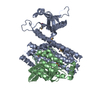
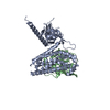
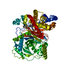
| Title | Crystal Structure of the Dbl and Pleckstrin homology domains of Dbs in complex with RhoA | ||||||
 Components Components |
| ||||||
 Keywords Keywords | SIGNALING PROTEIN / GUANINE NUCLEOTIDE EXCHANGE FACTOR / SMALL G-PROTEIN / RHOA / DBS / DH domain / PH domain | ||||||
| Function / homology |  Function and homology information Function and homology informationRHOC GTPase cycle / CDC42 GTPase cycle / NRAGE signals death through JNK / 1-phosphatidylinositol binding / RAC1 GTPase cycle / RHOB GTPase cycle / G alpha (12/13) signalling events / RHOA GTPase cycle / RHOG GTPase cycle / alpha-beta T cell lineage commitment ...RHOC GTPase cycle / CDC42 GTPase cycle / NRAGE signals death through JNK / 1-phosphatidylinositol binding / RAC1 GTPase cycle / RHOB GTPase cycle / G alpha (12/13) signalling events / RHOA GTPase cycle / RHOG GTPase cycle / alpha-beta T cell lineage commitment / aortic valve formation / positive regulation of lipase activity / endothelial tube lumen extension / skeletal muscle satellite cell migration / positive regulation of vascular associated smooth muscle contraction / angiotensin-mediated vasoconstriction involved in regulation of systemic arterial blood pressure / SLIT2:ROBO1 increases RHOA activity / RHO GTPases Activate Rhotekin and Rhophilins / Roundabout signaling pathway / bone trabecula morphogenesis / negative regulation of intracellular steroid hormone receptor signaling pathway / Axonal growth inhibition (RHOA activation) / Axonal growth stimulation / cleavage furrow formation / regulation of neural precursor cell proliferation / regulation of osteoblast proliferation / regulation of modification of postsynaptic actin cytoskeleton / forebrain radial glial cell differentiation / mitotic cleavage furrow formation / apical junction assembly / negative regulation of cell migration involved in sprouting angiogenesis / beta selection / cell junction assembly / establishment of epithelial cell apical/basal polarity / cellular response to chemokine / regulation of systemic arterial blood pressure by endothelin / negative regulation of oxidative phosphorylation / negative regulation of motor neuron apoptotic process / regulation of modification of postsynaptic structure / RHO GTPases Activate ROCKs / RHO GTPases activate CIT / negative regulation of cell size / Sema4D induced cell migration and growth-cone collapse / PCP/CE pathway / RHO GTPases activate KTN1 / positive regulation of alpha-beta T cell differentiation / positive regulation of podosome assembly / apolipoprotein A-I-mediated signaling pathway / wound healing, spreading of cells / Sema4D mediated inhibition of cell attachment and migration / positive regulation of leukocyte adhesion to vascular endothelial cell / motor neuron apoptotic process / odontogenesis / Wnt signaling pathway, planar cell polarity pathway / PI3K/AKT activation / ossification involved in bone maturation / regulation of focal adhesion assembly / androgen receptor signaling pathway / extrinsic component of cytoplasmic side of plasma membrane / negative chemotaxis / EPHA-mediated growth cone collapse / apical junction complex / stress fiber assembly / myosin binding / regulation of neuron projection development / positive regulation of cytokinesis / RHOC GTPase cycle / cellular response to cytokine stimulus / positive regulation of Rho protein signal transduction / cerebral cortex cell migration / ERBB2 Regulates Cell Motility / positive regulation of protein serine/threonine kinase activity / cleavage furrow / semaphorin-plexin signaling pathway / negative regulation of cell-substrate adhesion / ficolin-1-rich granule membrane / RHOA GTPase cycle / mitotic spindle assembly / positive regulation of T cell migration / endothelial cell migration / skeletal muscle tissue development / Rho protein signal transduction / RHO GTPases activate PKNs / GPVI-mediated activation cascade / PTK6 Regulates RHO GTPases, RAS GTPase and MAP kinases / negative regulation of reactive oxygen species biosynthetic process / positive regulation of stress fiber assembly / cytoplasmic microtubule organization / EPHB-mediated forward signaling / endomembrane system / positive regulation of neuron differentiation / substrate adhesion-dependent cell spreading / phosphatidylinositol binding / substantia nigra development / regulation of cell migration / guanyl-nucleotide exchange factor activity / secretory granule membrane / cell-matrix adhesion / regulation of microtubule cytoskeleton organization / cell periphery Similarity search - Function | ||||||
| Biological species |   Homo sapiens (human) Homo sapiens (human) | ||||||
| Method |  X-RAY DIFFRACTION / SYNCHROTRON / MOLECULAR REPLACEMENT / Resolution: 2.81 Å X-RAY DIFFRACTION / SYNCHROTRON / MOLECULAR REPLACEMENT / Resolution: 2.81 Å | ||||||
 Authors Authors | Snyder, J.T. / Worthylake, D.K. / Rossman, K.L. / Betts, L. / Pruitt, W.M. / Siderovski, D.P. / Der, C.J. / Sondek, J. | ||||||
 Citation Citation | Journal: Nat.Struct.Biol. / Year: 2002 Title: Structural basis for the selective activation of Rho GTPases by Dbl exchange factors. Authors: Snyder, J.T. / Worthylake, D.K. / Rossman, K.L. / Betts, L. / Pruitt, W.M. / Siderovski, D.P. / Der, C.J. / Sondek, J. | ||||||
| History |
|
- Structure visualization
| Structure viewer | Molecule: MolmilJmol/JSmol |
|---|
- Downloads & links
-Download
| PDBx/mmCIF format | 1lb1.cif.gz | 364.7 KB | Display | PDBx/mmCIF format |
|---|---|---|---|---|
| PDB format | pdb1lb1.ent.gz | 305.8 KB | Display | PDB format |
| PDBx/mmJSON format | 1lb1.json.gz | Tree view | PDBx/mmJSON format | |
| Others |  Other downloads Other downloads |
-Validation report
| Arichive directory | https://data.pdbj.org/pub/pdb/validation_reports/lb/1lb1ftp://data.pdbj.org/pub/pdb/validation_reports/lb/1lb1 | HTTPS FTP |
|---|
-Related structure data
| Related structure data | 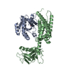 1ki1C 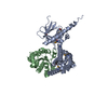 1kz7S S: Starting model for refinement C: citing same article ( |
|---|---|
| Similar structure data |


-Links
 PDBj
PDBj


- Assembly
| Deposited unit | 
| ||||||||
|---|---|---|---|---|---|---|---|---|---|
| 1 | 
| ||||||||
| 2 | 
| ||||||||
| 3 | 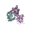
| ||||||||
| 4 | 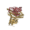
| ||||||||
| Unit cell |
|
-Components
| #1: Protein | Mass: 41252.332 Da / Num. of mol.: 4 Fragment: DBL homology domain (residues 623-818) and pleckstrin homology domain (residues 819-967) Source method: isolated from a genetically manipulated source Source: (gene. exp.)  #2: Protein | Mass: 21585.775 Da / Num. of mol.: 4 / Fragment: residues 1-190 / Mutation: C190S Source method: isolated from a genetically manipulated source Source: (gene. exp.) Homo sapiens (human) / Gene: RhoA / Plasmid: pPro EX HT / Species (production host): Escherichia coli / Production host: |
|---|
-Experimental details
-Experiment
| Experiment | Method: X-RAY DIFFRACTION / Number of used crystals: 1 |
|---|
- Sample preparation
| Crystal | Density Matthews: 3.81 Å3/Da / Density % sol: 67.72 % | |||||||||||||||||||||||||||||||||||||||||||||||||||||||||||||||
|---|---|---|---|---|---|---|---|---|---|---|---|---|---|---|---|---|---|---|---|---|---|---|---|---|---|---|---|---|---|---|---|---|---|---|---|---|---|---|---|---|---|---|---|---|---|---|---|---|---|---|---|---|---|---|---|---|---|---|---|---|---|---|---|---|
| Crystal grow | Temperature: 292 K / Method: vapor diffusion, sitting drop / pH: 8.4 Details: PEG 3350, lithium citrate, Inositol 1,4,5-trisphosphate, pH 8.4, VAPOR DIFFUSION, SITTING DROP, temperature 292K | |||||||||||||||||||||||||||||||||||||||||||||||||||||||||||||||
| Crystal grow | *PLUS Temperature: 18 ℃ / pH: 8 / Method: vapor diffusion | |||||||||||||||||||||||||||||||||||||||||||||||||||||||||||||||
| Components of the solutions | *PLUS
|
-Data collection
| Diffraction | Mean temperature: 80 K |
|---|---|
| Diffraction source | Source: SYNCHROTRON / Site: APS  / Beamline: 19-BM / Wavelength: 0.9765 Å / Beamline: 19-BM / Wavelength: 0.9765 Å |
| Detector | Type: ADSC QUANTUM 4 / Detector: CCD / Date: Feb 15, 2002 |
| Radiation | Protocol: SINGLE WAVELENGTH / Monochromatic (M) / Laue (L): M / Scattering type: x-ray |
| Radiation wavelength | Wavelength: 0.9765 Å / Relative weight: 1 |
| Reflection | Resolution: 2.81→47.7 Å / Num. all: 85993 / Num. obs: 85993 / Observed criterion σ(F): 0 / Observed criterion σ(I): -3 / Biso Wilson estimate: 87.9 Å2 / Limit h max: 56 / Limit h min: 0 / Limit k max: 56 / Limit k min: 0 / Limit l max: 53 / Limit l min: 0 / Observed criterion F max: 2986395.32 / Observed criterion F min: 8.8 |
| Reflection | *PLUS Highest resolution: 2.8 Å / Lowest resolution: 50 Å / Num. obs: 86038 / % possible obs: 99.1 % / Num. measured all: 269910 / Rmerge(I) obs: 0.078 |
| Reflection shell | *PLUS % possible obs: 94.1 % / Rmerge(I) obs: 0.654 / Mean I/σ(I) obs: 1.5 |
- Processing
| Software |
| ||||||||||||||||||||||||||||||||||||||||||||||||||||||||||||||||||||||||||||||||||||||||||
|---|---|---|---|---|---|---|---|---|---|---|---|---|---|---|---|---|---|---|---|---|---|---|---|---|---|---|---|---|---|---|---|---|---|---|---|---|---|---|---|---|---|---|---|---|---|---|---|---|---|---|---|---|---|---|---|---|---|---|---|---|---|---|---|---|---|---|---|---|---|---|---|---|---|---|---|---|---|---|---|---|---|---|---|---|---|---|---|---|---|---|---|
| Refinement | Method to determine structure: MOLECULAR REPLACEMENT Starting model: Dbs-Cdc42 (pdb code 1KZ7) Resolution: 2.81→47.7 Å / Rfactor Rfree error: 0.004 / Occupancy max: 1 / Occupancy min: 1 / Cross valid method: THROUGHOUT / σ(F): 0
| ||||||||||||||||||||||||||||||||||||||||||||||||||||||||||||||||||||||||||||||||||||||||||
| Solvent computation | Solvent model: CNS bulk solvent model used / Bsol: 45.4826 Å2 / ksol: 0.33567 e/Å3 | ||||||||||||||||||||||||||||||||||||||||||||||||||||||||||||||||||||||||||||||||||||||||||
| Displacement parameters | Biso max: 200.54 Å2 / Biso mean: 80.6 Å2 / Biso min: 4.81 Å2
| ||||||||||||||||||||||||||||||||||||||||||||||||||||||||||||||||||||||||||||||||||||||||||
| Refine analyze |
| ||||||||||||||||||||||||||||||||||||||||||||||||||||||||||||||||||||||||||||||||||||||||||
| Refinement step | Cycle: LAST / Resolution: 2.81→47.7 Å
| ||||||||||||||||||||||||||||||||||||||||||||||||||||||||||||||||||||||||||||||||||||||||||
| Refine LS restraints |
| ||||||||||||||||||||||||||||||||||||||||||||||||||||||||||||||||||||||||||||||||||||||||||
| LS refinement shell | Refine-ID: X-RAY DIFFRACTION
| ||||||||||||||||||||||||||||||||||||||||||||||||||||||||||||||||||||||||||||||||||||||||||
| Xplor file |
| ||||||||||||||||||||||||||||||||||||||||||||||||||||||||||||||||||||||||||||||||||||||||||
| Software | *PLUS Name: CNS / Classification: refinement | ||||||||||||||||||||||||||||||||||||||||||||||||||||||||||||||||||||||||||||||||||||||||||
| Refinement | *PLUS Highest resolution: 2.8 Å / Lowest resolution: 50 Å / Rfactor obs: 0.236 / Rfactor Rfree: 0.266 / Rfactor Rwork: 0.236 | ||||||||||||||||||||||||||||||||||||||||||||||||||||||||||||||||||||||||||||||||||||||||||
| Solvent computation | *PLUS | ||||||||||||||||||||||||||||||||||||||||||||||||||||||||||||||||||||||||||||||||||||||||||
| Displacement parameters | *PLUS | ||||||||||||||||||||||||||||||||||||||||||||||||||||||||||||||||||||||||||||||||||||||||||
| Refine LS restraints | *PLUS
| ||||||||||||||||||||||||||||||||||||||||||||||||||||||||||||||||||||||||||||||||||||||||||
| LS refinement shell | *PLUS Rfactor Rfree: 0.437 / Rfactor Rwork: 0.403 / Rfactor obs: 0.403 |
