 Movie
Movie Controller

[English] 日本語
 Yorodumi
- PDB-1d1u: USE OF AN N-TERMINAL FRAGMENT FROM MOLONEY MURINE LEUKEMIA VIRUS ... -
+ Open data

- Basic information
| Entry | Database: PDB / ID: 1d1u | ||||||
|---|---|---|---|---|---|---|---|
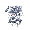
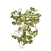
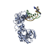
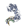
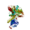
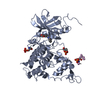
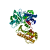
| Title | USE OF AN N-TERMINAL FRAGMENT FROM MOLONEY MURINE LEUKEMIA VIRUS REVERSE TRANSCRIPTASE TO FACILITATE CRYSTALLIZATION AND ANALYSIS OF A PSEUDO-16-MER DNA MOLECULE CONTAINING G-A MISPAIRS | ||||||
 Components Components |
| ||||||
 Keywords Keywords | HYDROLASE/DNA / G-A MISPAIR / SYN-ADENINE / NUCLEIC ACID / PROTEIN-DNA COMPLEX / SINGLE-STRAND OVERHANG / REVERSE TRANSCRIPTASE / MOLONEY MURINE LEUKEMIA VIRUS / HYDROLASE-DNA COMPLEX | ||||||
| Function / homology |  Function and homology information Function and homology informationretroviral 3' processing activity / host cell late endosome membrane / DNA catabolic process / Hydrolases; Acting on peptide bonds (peptidases); Aspartic endopeptidases / ribonuclease H / virion assembly / protein-DNA complex / viral genome integration into host DNA / establishment of integrated proviral latency / RNA-directed DNA polymerase ...retroviral 3' processing activity / host cell late endosome membrane / DNA catabolic process / Hydrolases; Acting on peptide bonds (peptidases); Aspartic endopeptidases / ribonuclease H / virion assembly / protein-DNA complex / viral genome integration into host DNA / establishment of integrated proviral latency / RNA-directed DNA polymerase / host multivesicular body / RNA-directed DNA polymerase activity / RNA-DNA hybrid ribonuclease activity / Transferases; Transferring phosphorus-containing groups; Nucleotidyltransferases / viral nucleocapsid / DNA recombination / DNA-directed DNA polymerase / structural constituent of virion / aspartic-type endopeptidase activity / Hydrolases; Acting on ester bonds / DNA-directed DNA polymerase activity / symbiont-mediated suppression of host gene expression / symbiont entry into host cell / host cell plasma membrane / proteolysis / DNA binding / RNA binding / zinc ion binding Similarity search - Function | ||||||
| Biological species |  Moloney murine leukemia virus Moloney murine leukemia virus | ||||||
| Method |  X-RAY DIFFRACTION / Resolution: 2.3 Å X-RAY DIFFRACTION / Resolution: 2.3 Å | ||||||
 Authors Authors | Cote, M.L. / Yohannan, S. / Georgiadis, M.M. | ||||||
 Citation Citation | Journal: Acta Crystallogr.,Sect.D / Year: 2000 Title: Use of an N-terminal fragment from moloney murine leukemia virus reverse transcriptase to facilitate crystallization and analysis of a pseudo-16-mer DNA molecule containing G-A mispairs. Authors: Cote, M.L. / Yohannan, S.J. / Georgiadis, M.M. #1: Journal: J.Mol.Biol. / Year: 2000Title: Crystal Structures of the N-Terminal Fragment from Moloney Murine Leukemia Virus Reverse Transcriptase Complexed with Nucleic Acid: Functional Implications for Template-Primer Binding to the Fingers Domain Authors: Najmudin, S. / Cote, M.L. / Sun, D. / Yohannan, S. / Montano, S.P. / Gu, J. / Georgiadis, M.M. | ||||||
| History |
|
- Structure visualization
| Structure viewer | Molecule: MolmilJmol/JSmol |
|---|
- Downloads & links
-Download
| PDBx/mmCIF format | 1d1u.cif.gz | 79.8 KB | Display | PDBx/mmCIF format |
|---|---|---|---|---|
| PDB format | pdb1d1u.ent.gz | 56.6 KB | Display | PDB format |
| PDBx/mmJSON format | 1d1u.json.gz | Tree view | PDBx/mmJSON format | |
| Others |  Other downloads Other downloads |
-Validation report
| Arichive directory | https://data.pdbj.org/pub/pdb/validation_reports/d1/1d1uftp://data.pdbj.org/pub/pdb/validation_reports/d1/1d1u | HTTPS FTP |
|---|
-Related structure data
| Similar structure data |
|---|


-Links
 PDBj
PDBj

- Assembly
| Deposited unit | 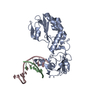
| ||||||||
|---|---|---|---|---|---|---|---|---|---|
| 1 |
| ||||||||
| Unit cell |
|
-Components
| #1: DNA chain | Mass: 1800.203 Da / Num. of mol.: 1 / Source method: obtained synthetically / Details: SYNTHETIC |
|---|---|
| #2: DNA chain | Mass: 3079.031 Da / Num. of mol.: 1 / Source method: obtained synthetically Details: THE SEQUENCE 5'-CTCGTG-3' WAS SYNTHESIZED ON AN APPLIED BIOSYSTEMS 392 DNA/RNA SYNTHESIZER |
| #3: Protein | Mass: 28934.287 Da / Num. of mol.: 1 / Fragment: FINGERS AND PALM DOMAIN OF MMLV RT Source method: isolated from a genetically manipulated source Source: (gene. exp.) Moloney murine leukemia virus / Genus: Gammaretrovirus / Species: Murine leukemia virus / Gene: MMLV REVERSE TRANSCRIPTASE / Production host:  |
| #4: Water | ChemComp-HOH /  Mass: 18.015 Da / Num. of mol.: 175 / Source method: isolated from a natural source / Formula: H2O Mass: 18.015 Da / Num. of mol.: 175 / Source method: isolated from a natural source / Formula: H2O |
-Experimental details
-Experiment
| Experiment | Method: X-RAY DIFFRACTION / Number of used crystals: 1 |
|---|
- Sample preparation
| Crystal | Density Matthews: 2.73 Å3/Da / Density % sol: 54.98 % | ||||||||||||||||||||||||||||||||||||||||||
|---|---|---|---|---|---|---|---|---|---|---|---|---|---|---|---|---|---|---|---|---|---|---|---|---|---|---|---|---|---|---|---|---|---|---|---|---|---|---|---|---|---|---|---|
| Crystal grow | pH: 6.5 / Details: pH 6.50 | ||||||||||||||||||||||||||||||||||||||||||
| Crystal grow | *PLUS Temperature: 293 K / Method: vapor diffusion, hanging drop | ||||||||||||||||||||||||||||||||||||||||||
| Components of the solutions | *PLUS
|
-Data collection
| Diffraction | Mean temperature: 108 K |
|---|---|
| Diffraction source | Source: ROTATING ANODE / Type: RIGAKU / Wavelength: 1.5418 |
| Detector | Type: RIGAKU RAXIS IV / Detector: IMAGE PLATE / Date: Jun 1, 1998 |
| Radiation | Protocol: SINGLE WAVELENGTH / Monochromatic (M) / Laue (L): M / Scattering type: x-ray |
| Radiation wavelength | Wavelength: 1.5418 Å / Relative weight: 1 |
| Reflection | Resolution: 2.3→50 Å / Num. obs: 17117 / % possible obs: 98.9 % / Observed criterion σ(I): 0 / Redundancy: 5.5 % / Biso Wilson estimate: 41.3 Å2 / Rmerge(I) obs: 0.069 / Net I/σ(I): 22.8 |
| Reflection shell | Resolution: 2.3→2.38 Å / Redundancy: 3.3 % / Rmerge(I) obs: 0.278 / % possible all: 98.5 |
| Reflection shell | *PLUS % possible obs: 98.5 % / Mean I/σ(I) obs: 4.6 |
- Processing
| Software |
| ||||||||||||||||||||||||||||||||||||||||||||||||||||||||||||
|---|---|---|---|---|---|---|---|---|---|---|---|---|---|---|---|---|---|---|---|---|---|---|---|---|---|---|---|---|---|---|---|---|---|---|---|---|---|---|---|---|---|---|---|---|---|---|---|---|---|---|---|---|---|---|---|---|---|---|---|---|---|
| Refinement | Resolution: 2.3→50 Å / σ(F): 0 / Stereochemistry target values: ENGH & HUBER Details: MODEL WAS REFINED WITH TWO DIFFERENT SIDE-CHAIN CONFORMATIONS FOR TYR 64 (IN THE A MOLECULE). BOTH WERE HELD AT 50% OCCUPANCY. THE 5'- TERMINAL ADENINE OF THE "C" CHAIN MAY HAVE EITHER THE ...Details: MODEL WAS REFINED WITH TWO DIFFERENT SIDE-CHAIN CONFORMATIONS FOR TYR 64 (IN THE A MOLECULE). BOTH WERE HELD AT 50% OCCUPANCY. THE 5'- TERMINAL ADENINE OF THE "C" CHAIN MAY HAVE EITHER THE SYN- OR ANTI- CONFORMATION. THE DEPOSITED DATA HAVE ADENINE C7 IN THE ANTI- CONFORMATION. INDEPENDENT REFINEMENTS OF THE STRUCTURAL MODEL WITH EITHER A SYN- OR AN ANTI-CONFORMATION FOR THE 5'-TERMINAL ADENINE ((A7) IN THE "C" CHAIN) YIELDED ELECTRON DENSITY MAPS WHICH GAVE NO PREFERENCE FOR ONE CONFORMATION OVER THE OTHER, AND NO INDICATION OF PARTIAL OCCUPANCIES FOR BOTH. THE INDEPENDENT REFINEMENTS YIELDED THE SAME REFINEMENT STATISTICS. THE DEPOSITED DATA CONTAIN THE ADENINE (A7) IN THE ANTI-CONFORMATION. THE COORDINATES FOR THE SYN-ADENINE ARE AVAILABLE UPON REQUEST FROM THE AUTHORS
| ||||||||||||||||||||||||||||||||||||||||||||||||||||||||||||
| Refine analyze |
| ||||||||||||||||||||||||||||||||||||||||||||||||||||||||||||
| Refinement step | Cycle: LAST / Resolution: 2.3→50 Å
| ||||||||||||||||||||||||||||||||||||||||||||||||||||||||||||
| Refine LS restraints |
| ||||||||||||||||||||||||||||||||||||||||||||||||||||||||||||
| Software | *PLUS Name: CNS / Classification: refinement | ||||||||||||||||||||||||||||||||||||||||||||||||||||||||||||
| Refine LS restraints | *PLUS
|
