 Movie
Movie Controller

[English] 日本語
 Yorodumi
- PDB-1i6j: CRYSTAL STRUCTURE OF A PSEUDO-16-MER DNA WITH STACKED GUANINES AN... -
+ Open data

- Basic information
| Entry | Database: PDB / ID: 1i6j | ||||||
|---|---|---|---|---|---|---|---|
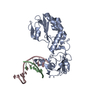
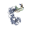
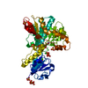
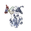
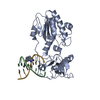
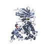
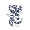
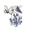
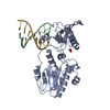
| Title | CRYSTAL STRUCTURE OF A PSEUDO-16-MER DNA WITH STACKED GUANINES AND TWO G-A MISPAIRS COMPLEXED WITH THE N-TERMINAL FRAGMENT OF MOLONEY MURINE LEUKEMIA VIRUS REVERSE TRANSCRIPTASE | ||||||
 Components Components |
| ||||||
 Keywords Keywords | TRANSFERASE/DNA / Moloney Murine Leukemia Virus / Reverse Transcriptase / G-A Mispair / TRANSFERASE-DNA COMPLEX | ||||||
| Function / homology |  Function and homology information Function and homology informationretroviral 3' processing activity / host cell late endosome membrane / DNA catabolic process / Hydrolases; Acting on peptide bonds (peptidases); Aspartic endopeptidases / ribonuclease H / virion assembly / protein-DNA complex / viral genome integration into host DNA / establishment of integrated proviral latency / RNA-directed DNA polymerase ...retroviral 3' processing activity / host cell late endosome membrane / DNA catabolic process / Hydrolases; Acting on peptide bonds (peptidases); Aspartic endopeptidases / ribonuclease H / virion assembly / protein-DNA complex / viral genome integration into host DNA / establishment of integrated proviral latency / RNA-directed DNA polymerase / host multivesicular body / RNA-directed DNA polymerase activity / RNA-DNA hybrid ribonuclease activity / Transferases; Transferring phosphorus-containing groups; Nucleotidyltransferases / viral nucleocapsid / DNA recombination / DNA-directed DNA polymerase / structural constituent of virion / aspartic-type endopeptidase activity / Hydrolases; Acting on ester bonds / DNA-directed DNA polymerase activity / symbiont-mediated suppression of host gene expression / symbiont entry into host cell / host cell plasma membrane / proteolysis / DNA binding / RNA binding / zinc ion binding Similarity search - Function | ||||||
| Biological species |  Moloney murine leukemia virus Moloney murine leukemia virus | ||||||
| Method |  X-RAY DIFFRACTION / MOLECULAR REPLACEMENT / Resolution: 2 Å X-RAY DIFFRACTION / MOLECULAR REPLACEMENT / Resolution: 2 Å | ||||||
 Authors Authors | Cote, M.L. / Georgiadis, M.M. | ||||||
 Citation Citation | Journal: Acta Crystallogr.,Sect.D / Year: 2001 Title: Structure of a pseudo-16-mer DNA with stacked guanines and two G-A mispairs complexed with the N-terminal fragment of Moloney murine leukemia virus reverse transcriptase. Authors: Cote, M.L. / Georgiadis, M.M. | ||||||
| History |
|
- Structure visualization
| Structure viewer | Molecule: MolmilJmol/JSmol |
|---|
- Downloads & links
-Download
| PDBx/mmCIF format | 1i6j.cif.gz | 79.8 KB | Display | PDBx/mmCIF format |
|---|---|---|---|---|
| PDB format | pdb1i6j.ent.gz | 55.8 KB | Display | PDB format |
| PDBx/mmJSON format | 1i6j.json.gz | Tree view | PDBx/mmJSON format | |
| Others |  Other downloads Other downloads |
-Validation report
| Arichive directory | https://data.pdbj.org/pub/pdb/validation_reports/i6/1i6jftp://data.pdbj.org/pub/pdb/validation_reports/i6/1i6j | HTTPS FTP |
|---|
-Related structure data
| Related structure data |  1d1uS S: Starting model for refinement |
|---|---|
| Similar structure data |

-Links
 PDBj
PDBj

- Assembly
| Deposited unit | 
| ||||||||
|---|---|---|---|---|---|---|---|---|---|
| 1 |
| ||||||||
| Unit cell |
| ||||||||
| Details | The second part of the DNA is generated by the 2-fold: -x-1, -y, z |
-Components
| #1: DNA chain | Mass: 1800.203 Da / Num. of mol.: 1 / Source method: obtained synthetically / Details: ABI 392 DNA Synthesizer |
|---|---|
| #2: DNA chain | Mass: 3079.031 Da / Num. of mol.: 1 / Source method: obtained synthetically / Details: ABI 392 DNA Synthesizer |
| #3: Protein | Mass: 29065.480 Da / Num. of mol.: 1 / Fragment: N-TERMINAL FRAGMENT Source method: isolated from a genetically manipulated source Source: (gene. exp.) Moloney murine leukemia virus / Genus: Gammaretrovirus / Species: Murine leukemia virus / Gene: VIRUS / Plasmid: PET15B / Species (production host): Escherichia coli / Production host:  |
| #4: Water | ChemComp-HOH /  Mass: 18.015 Da / Num. of mol.: 204 / Source method: isolated from a natural source / Formula: H2O Mass: 18.015 Da / Num. of mol.: 204 / Source method: isolated from a natural source / Formula: H2O |
-Experimental details
-Experiment
| Experiment | Method: X-RAY DIFFRACTION / Number of used crystals: 1 |
|---|
- Sample preparation
| Crystal | Density Matthews: 2.72 Å3/Da / Density % sol: 54.74 % | ||||||||||||||||||||||||||||||||||||
|---|---|---|---|---|---|---|---|---|---|---|---|---|---|---|---|---|---|---|---|---|---|---|---|---|---|---|---|---|---|---|---|---|---|---|---|---|---|
| Crystal grow | Temperature: 277 K / Method: vapor diffusion, hanging drop / pH: 6.5 Details: PEG 4000, NaCl, ADA, MgCl2, HEPES, pH 6.5, VAPOR DIFFUSION, HANGING DROP, temperature 277K | ||||||||||||||||||||||||||||||||||||
| Components of the solutions |
| ||||||||||||||||||||||||||||||||||||
| Crystal grow | *PLUS Temperature: 293 K | ||||||||||||||||||||||||||||||||||||
| Components of the solutions | *PLUS
|
-Data collection
| Diffraction | Mean temperature: 108 K |
|---|---|
| Diffraction source | Source: ROTATING ANODE / Type: RIGAKU / Wavelength: 1.5418 Å |
| Detector | Type: RIGAKU RAXIS IV / Detector: IMAGE PLATE / Date: Feb 28, 2000 / Details: mirrors |
| Radiation | Monochromator: Ni FILTER / Protocol: SINGLE WAVELENGTH / Monochromatic (M) / Laue (L): M / Scattering type: x-ray |
| Radiation wavelength | Wavelength: 1.5418 Å / Relative weight: 1 |
| Reflection | Resolution: 2→50 Å / Num. all: 25995 / Num. obs: 24781 / % possible obs: 95.3 % / Observed criterion σ(F): 0 / Observed criterion σ(I): 0 / Redundancy: 1.1 % / Biso Wilson estimate: 41.3 Å2 / Rmerge(I) obs: 0.028 / Net I/σ(I): 28.2 |
| Reflection shell | Resolution: 2→2.07 Å / Redundancy: 2.1 % / Rmerge(I) obs: 0.298 / Mean I/σ(I) obs: 3.4 / Num. unique all: 2163 / % possible all: 85.4 |
| Reflection | *PLUS Highest resolution: 2 Å / Lowest resolution: 50 Å |
| Reflection shell | *PLUS % possible obs: 85.4 % |
- Processing
| Software |
| |||||||||||||||||||||||||
|---|---|---|---|---|---|---|---|---|---|---|---|---|---|---|---|---|---|---|---|---|---|---|---|---|---|---|
| Refinement | Method to determine structure: MOLECULAR REPLACEMENT Starting model: PDB ENTRY 1D1U Resolution: 2→50 Å / Isotropic thermal model: Isotropic / Cross valid method: THROUGHOUT / σ(F): 0 / σ(I): 0 / Stereochemistry target values: Engh & Huber
| |||||||||||||||||||||||||
| Displacement parameters | Biso mean: 44 Å2 | |||||||||||||||||||||||||
| Refine analyze | Luzzati coordinate error obs: 0.28 Å / Luzzati sigma a obs: 0.2 Å | |||||||||||||||||||||||||
| Refinement step | Cycle: LAST / Resolution: 2→50 Å
| |||||||||||||||||||||||||
| Refine LS restraints |
| |||||||||||||||||||||||||
| Software | *PLUS Name: CNS / Version: 1 / Classification: refinement | |||||||||||||||||||||||||
| Refinement | *PLUS σ(F): 0 / Rfactor obs: 0.225 | |||||||||||||||||||||||||
| Solvent computation | *PLUS | |||||||||||||||||||||||||
| Displacement parameters | *PLUS Biso mean: 44 Å2 | |||||||||||||||||||||||||
| Refine LS restraints | *PLUS
|
