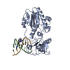
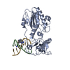
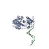
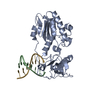
Two protein molecules joined by a 16mer-bp double strand of DNA, with CD27 bound to both 5'-AATT sites. One asymmetric unit retains one half of the 16mer-bp, one protein and one bound ligand.
-
Components
-
Protein , 1 types, 1 molecules A
#1: Protein
Reversetranscriptasedomain / RT
Mass: 28934.287 Da / Num. of mol.: 1 Fragment: Reverse transcriptase domain: UNP residues 144-398 Source method: isolated from a genetically manipulated source Source: (gene. exp.) Moloney murine leukemia virus / Gene: MoMLV, pol / Plasmid: pET15b / Production host: Escherichia coli (E. coli) / Strain (production host): BL21 Rosetta / References: UniProt: P03355, RNA-directed DNA polymerase
-
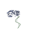
DNA chain , 2 types, 2 molecules GB
#2: DNA chain
5'-D(*CP*TP*TP*AP*AP*TP*TP*C)-3'
Mass: 2376.591 Da / Num. of mol.: 1 / Source method: obtained synthetically
#3: DNA chain
5'-D(P*GP*AP*AP*TP*TP*AP*AP*G)-3'
Mass: 2474.667 Da / Num. of mol.: 1 / Source method: obtained synthetically
In the structure databanks used in Yorodumi, some data are registered as the other names, "COVID-19 virus" and "2019-nCoV". Here are the details of the virus and the list of structure data.
Jan 31, 2019. EMDB accession codes are about to change! (news from PDBe EMDB page)
EMDB accession codes are about to change! (news from PDBe EMDB page)
The allocation of 4 digits for EMDB accession codes will soon come to an end. Whilst these codes will remain in use, new EMDB accession codes will include an additional digit and will expand incrementally as the available range of codes is exhausted. The current 4-digit format prefixed with “EMD-” (i.e. EMD-XXXX) will advance to a 5-digit format (i.e. EMD-XXXXX), and so on. It is currently estimated that the 4-digit codes will be depleted around Spring 2019, at which point the 5-digit format will come into force.
The EM Navigator/Yorodumi systems omit the EMD- prefix.
Related info.:Q: What is EMD? / ID/Accession-code notation in Yorodumi/EM Navigator
Yorodumi is a browser for structure data from EMDB, PDB, SASBDB, etc.
This page is also the successor to EM Navigator detail page, and also detail information page/front-end page for Omokage search.
The word "yorodu" (or yorozu) is an old Japanese word meaning "ten thousand". "mi" (miru) is to see.
Related info.:EMDB / PDB / SASBDB / Comparison of 3 databanks / Yorodumi Search / Aug 31, 2016. New EM Navigator & Yorodumi / Yorodumi Papers / Jmol/JSmol / Function and homology information / Changes in new EM Navigator and Yorodumi
 Movie
Movie Controller

 Yorodumi
 Open data
 Basic information
 Components
 Keywords
 Function and homology information
 Moloney murine leukemia virus
 X-RAY DIFFRACTION /
  Authors
 Citation
 Structure visualization
 Downloads & links
 Other downloads
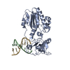


 PDBj
PDBj
 Assembly
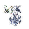


 Mass: 59.044 Da / Num. of mol.: 5 / Source method: obtained synthetically / Formula: C2H3O2
 Mass: 335.406 Da / Num. of mol.: 1 / Source method: obtained synthetically / Formula: C18H21N7
 Sample preparation
 / Beamline: 19-ID / Wavelength: 1.008001 Å
 Processing