 Movie
Movie Controller

[English] 日本語
 Yorodumi
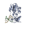
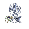
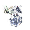
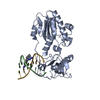
- PDB-6b1q: Hydrogen Bonding Complementary, not size complementarity is key i... -
+ Open data

- Basic information
| Entry | Database: PDB / ID: 6b1q | ||||||
|---|---|---|---|---|---|---|---|
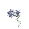
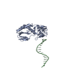
| Title | Hydrogen Bonding Complementary, not size complementarity is key in the formation of the double helix | ||||||
 Components Components |
| ||||||
 Keywords Keywords | DNA BINDING PROTEIN/DNA / Protein-DNA / AEGIS / unnatural base pair / host-guest system / DNA BINDING PROTEIN / DNA BINDING PROTEIN-DNA complex | ||||||
| Function / homology |  Function and homology information Function and homology informationretroviral 3' processing activity / host cell late endosome membrane / DNA catabolic process / Hydrolases; Acting on peptide bonds (peptidases); Aspartic endopeptidases / ribonuclease H / virion assembly / protein-DNA complex / viral genome integration into host DNA / establishment of integrated proviral latency / RNA-directed DNA polymerase ...retroviral 3' processing activity / host cell late endosome membrane / DNA catabolic process / Hydrolases; Acting on peptide bonds (peptidases); Aspartic endopeptidases / ribonuclease H / virion assembly / protein-DNA complex / viral genome integration into host DNA / establishment of integrated proviral latency / RNA-directed DNA polymerase / host multivesicular body / RNA-directed DNA polymerase activity / RNA-DNA hybrid ribonuclease activity / Transferases; Transferring phosphorus-containing groups; Nucleotidyltransferases / viral nucleocapsid / DNA recombination / DNA-directed DNA polymerase / structural constituent of virion / aspartic-type endopeptidase activity / Hydrolases; Acting on ester bonds / DNA-directed DNA polymerase activity / symbiont-mediated suppression of host gene expression / symbiont entry into host cell / host cell plasma membrane / proteolysis / DNA binding / RNA binding / zinc ion binding Similarity search - Function | ||||||
| Biological species |  Moloney murine leukemia virus Moloney murine leukemia virus | ||||||
| Method |  X-RAY DIFFRACTION / SYNCHROTRON / MOLECULAR REPLACEMENT / molecular replacement / Resolution: 1.9 Å X-RAY DIFFRACTION / SYNCHROTRON / MOLECULAR REPLACEMENT / molecular replacement / Resolution: 1.9 Å | ||||||
 Authors Authors | Singh, I. / Georgiadis, M.M. | ||||||
 Citation Citation | Journal: J. Am. Chem. Soc. / Year: 2018 Title: "Skinny" and "Fat" DNA: Two New Double Helices. Authors: Hoshika, S. / Singh, I. / Switzer, C. / Molt Jr., R.W. / Leal, N.A. / Kim, M.J. / Kim, M.S. / Kim, H.J. / Georgiadis, M.M. / Benner, S.A. | ||||||
| History |
|
- Structure visualization
| Structure viewer | Molecule: MolmilJmol/JSmol |
|---|
- Downloads & links
-Download
| PDBx/mmCIF format | 6b1q.cif.gz | 84.4 KB | Display | PDBx/mmCIF format |
|---|---|---|---|---|
| PDB format | pdb6b1q.ent.gz | 58.4 KB | Display | PDB format |
| PDBx/mmJSON format | 6b1q.json.gz | Tree view | PDBx/mmJSON format | |
| Others |  Other downloads Other downloads |
-Validation report
| Arichive directory | https://data.pdbj.org/pub/pdb/validation_reports/b1/6b1qftp://data.pdbj.org/pub/pdb/validation_reports/b1/6b1q | HTTPS FTP |
|---|
-Related structure data
| Related structure data |  6b1rC  6b1sC  4xo0S S: Starting model for refinement C: citing same article ( |
|---|---|
| Similar structure data |


-Links
 PDBj
PDBj
- Assembly
| Deposited unit | 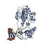
| ||||||||
|---|---|---|---|---|---|---|---|---|---|
| 1 | 
| ||||||||
| Unit cell |
|
-Components
| #1: Protein | Mass: 29347.754 Da / Num. of mol.: 1 / Fragment: Catalytic fragment (UNP residues 683-937) Source method: isolated from a genetically manipulated source Source: (gene. exp.) Moloney murine leukemia virus / Production host: |
|---|---|
| #2: DNA chain | Mass: 2457.618 Da / Num. of mol.: 1 / Source method: obtained synthetically / Source: (synth.) |
| #3: DNA chain | Mass: 2512.724 Da / Num. of mol.: 1 / Source method: obtained synthetically / Source: (synth.) |
| #4: Water | ChemComp-HOH /  Mass: 18.015 Da / Num. of mol.: 274 / Source method: isolated from a natural source / Formula: H2O Mass: 18.015 Da / Num. of mol.: 274 / Source method: isolated from a natural source / Formula: H2O |
-Experimental details
-Experiment
| Experiment | Method: X-RAY DIFFRACTION / Number of used crystals: 1 |
|---|
- Sample preparation
| Crystal | Density Matthews: 2.78 Å3/Da / Density % sol: 55.78 % |
|---|---|
| Crystal grow | Temperature: 277.15 K / Method: vapor diffusion, hanging drop / pH: 6.5 Details: 6 % PEG 4000, 5 mM magnesium acetate and 50 mM ADA (pH 6.5) |
-Data collection
| Diffraction | Mean temperature: 100 K | |||||||||||||||||||||||||||||||||||||||||||||||||||||||||||||||||||||||||||||||||||||||||||||||||||
|---|---|---|---|---|---|---|---|---|---|---|---|---|---|---|---|---|---|---|---|---|---|---|---|---|---|---|---|---|---|---|---|---|---|---|---|---|---|---|---|---|---|---|---|---|---|---|---|---|---|---|---|---|---|---|---|---|---|---|---|---|---|---|---|---|---|---|---|---|---|---|---|---|---|---|---|---|---|---|---|---|---|---|---|---|---|---|---|---|---|---|---|---|---|---|---|---|---|---|---|---|
| Diffraction source | Source: SYNCHROTRON / Site: APS  / Beamline: 31-ID / Wavelength: 0.97931 Å / Beamline: 31-ID / Wavelength: 0.97931 Å | |||||||||||||||||||||||||||||||||||||||||||||||||||||||||||||||||||||||||||||||||||||||||||||||||||
| Detector | Type: RAYONIX MX-225 / Detector: CCD / Date: Apr 15, 2016 | |||||||||||||||||||||||||||||||||||||||||||||||||||||||||||||||||||||||||||||||||||||||||||||||||||
| Radiation | Protocol: SINGLE WAVELENGTH / Monochromatic (M) / Laue (L): M / Scattering type: x-ray | |||||||||||||||||||||||||||||||||||||||||||||||||||||||||||||||||||||||||||||||||||||||||||||||||||
| Radiation wavelength | Wavelength: 0.97931 Å / Relative weight: 1 | |||||||||||||||||||||||||||||||||||||||||||||||||||||||||||||||||||||||||||||||||||||||||||||||||||
| Reflection | Resolution: 1.896→146.282 Å / Num. obs: 31032 / % possible obs: 99.8 % / Redundancy: 7 % / Biso Wilson estimate: 21.9 Å2 / Rmerge(I) obs: 0.088 / Rpim(I) all: 0.039 / Rrim(I) all: 0.104 / Rsym value: 0.088 / Net I/av σ(I): 6.7 / Net I/σ(I): 14.4 | |||||||||||||||||||||||||||||||||||||||||||||||||||||||||||||||||||||||||||||||||||||||||||||||||||
| Reflection shell | Diffraction-ID: 1
|
-Phasing
| Phasing | Method: molecular replacement |
|---|
- Processing
| Software |
| ||||||||||||||||||||||||||||||||||||||||||||||||||||||||||||||||||||||||||||||||||||
|---|---|---|---|---|---|---|---|---|---|---|---|---|---|---|---|---|---|---|---|---|---|---|---|---|---|---|---|---|---|---|---|---|---|---|---|---|---|---|---|---|---|---|---|---|---|---|---|---|---|---|---|---|---|---|---|---|---|---|---|---|---|---|---|---|---|---|---|---|---|---|---|---|---|---|---|---|---|---|---|---|---|---|---|---|---|
| Refinement | Method to determine structure: MOLECULAR REPLACEMENT Starting model: 4XO0 Resolution: 1.9→44.696 Å / SU ML: 0.2 / Cross valid method: NONE / σ(F): 1.35 / Phase error: 23.43
| ||||||||||||||||||||||||||||||||||||||||||||||||||||||||||||||||||||||||||||||||||||
| Solvent computation | Shrinkage radii: 0.9 Å / VDW probe radii: 1.11 Å | ||||||||||||||||||||||||||||||||||||||||||||||||||||||||||||||||||||||||||||||||||||
| Displacement parameters | Biso max: 89.05 Å2 / Biso mean: 31.3501 Å2 / Biso min: 9.24 Å2 | ||||||||||||||||||||||||||||||||||||||||||||||||||||||||||||||||||||||||||||||||||||
| Refinement step | Cycle: final / Resolution: 1.9→44.696 Å
| ||||||||||||||||||||||||||||||||||||||||||||||||||||||||||||||||||||||||||||||||||||
| Refine LS restraints |
| ||||||||||||||||||||||||||||||||||||||||||||||||||||||||||||||||||||||||||||||||||||
| LS refinement shell | Refine-ID: X-RAY DIFFRACTION / Rfactor Rfree error: 0 / Total num. of bins used: 11
|
