Movie
Movie Controller

[English] 日本語
 Yorodumi
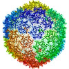
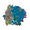
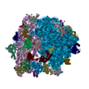
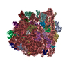
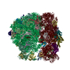
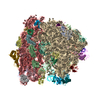
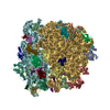
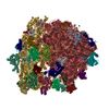
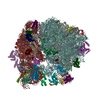
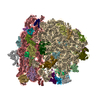
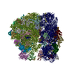
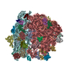
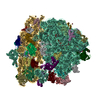
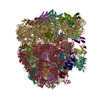
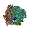
- PDB-4v61: Homology model for the Spinach chloroplast 30S subunit fitted to ... -
+ Open data

- Basic information
| Entry | Database: PDB / ID: 4v61 | |||||||||
|---|---|---|---|---|---|---|---|---|---|---|
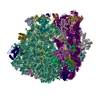
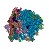
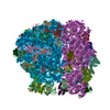
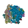
| Title | Homology model for the Spinach chloroplast 30S subunit fitted to 9.4A cryo-EM map of the 70S chlororibosome. | |||||||||
 Components Components |
| |||||||||
 Keywords Keywords | RIBOSOME / SMALL RIBOSOMAL SUBUNIT / SPINACH CHLOROPLAST RIBOSOME / RIBONUCLEOPROTEIN PARTICLE / MACROMOLECULAR COMPLEX | |||||||||
| Function / homology |  Function and homology information Function and homology informationplastid small ribosomal subunit / mitochondrial large ribosomal subunit / mitochondrial small ribosomal subunit / mitochondrial translation / chloroplast / DNA-templated transcription termination / large ribosomal subunit / transferase activity / ribosomal small subunit biogenesis / ribosomal small subunit assembly ...plastid small ribosomal subunit / mitochondrial large ribosomal subunit / mitochondrial small ribosomal subunit / mitochondrial translation / chloroplast / DNA-templated transcription termination / large ribosomal subunit / transferase activity / ribosomal small subunit biogenesis / ribosomal small subunit assembly / small ribosomal subunit / small ribosomal subunit rRNA binding / ribosomal large subunit assembly / cytosolic small ribosomal subunit / large ribosomal subunit rRNA binding / cytosolic large ribosomal subunit / negative regulation of translation / rRNA binding / structural constituent of ribosome / ribosome / translation / ribonucleoprotein complex / response to antibiotic / mRNA binding / mitochondrion / RNA binding Similarity search - Function | |||||||||
| Biological species |  Spinacea oleracea (spinach) Spinacea oleracea (spinach) | |||||||||
| Method | ELECTRON MICROSCOPY / single particle reconstruction / cryo EM / Resolution: 9.4 Å | |||||||||
 Authors Authors | Sharma, M.R. / Wilson, D.N. / Datta, P.P. / Barat, C. / Schluenzen, F. / Fucini, P. / Agrawal, R.K. | |||||||||
 Citation Citation | Journal: Proc Natl Acad Sci U S A / Year: 2007 Title: Cryo-EM study of the spinach chloroplast ribosome reveals the structural and functional roles of plastid-specific ribosomal proteins. Authors: Manjuli R Sharma / Daniel N Wilson / Partha P Datta / Chandana Barat / Frank Schluenzen / Paola Fucini / Rajendra K Agrawal /  Abstract: Protein synthesis in the chloroplast is carried out by chloroplast ribosomes (chloro-ribosome) and regulated in a light-dependent manner. Chloroplast or plastid ribosomal proteins (PRPs) generally ...Protein synthesis in the chloroplast is carried out by chloroplast ribosomes (chloro-ribosome) and regulated in a light-dependent manner. Chloroplast or plastid ribosomal proteins (PRPs) generally are larger than their bacterial counterparts, and chloro-ribosomes contain additional plastid-specific ribosomal proteins (PSRPs); however, it is unclear to what extent these proteins play structural or regulatory roles during translation. We have obtained a three-dimensional cryo-EM map of the spinach 70S chloro-ribosome, revealing the overall structural organization to be similar to bacterial ribosomes. Fitting of the conserved portions of the x-ray crystallographic structure of the bacterial 70S ribosome into our cryo-EM map of the chloro-ribosome reveals the positions of PRP extensions and the locations of the PSRPs. Surprisingly, PSRP1 binds in the decoding region of the small (30S) ribosomal subunit, in a manner that would preclude the binding of messenger and transfer RNAs to the ribosome, suggesting that PSRP1 is a translation factor rather than a ribosomal protein. PSRP2 and PSRP3 appear to structurally compensate for missing segments of the 16S rRNA within the 30S subunit, whereas PSRP4 occupies a position buried within the head of the 30S subunit. One of the two PSRPs in the large (50S) ribosomal subunit lies near the tRNA exit site. Furthermore, we find a mass of density corresponding to chloro-ribosome recycling factor; domain II of this factor appears to interact with the flexible C-terminal domain of PSRP1. Our study provides evolutionary insights into the structural and functional roles that the PSRPs play during protein synthesis in chloroplasts. | |||||||||
| History |
|
- Structure visualization
| Movie |
Movie viewer |
|---|---|
| Structure viewer | Molecule: MolmilJmol/JSmol |
- Downloads & links
-Download
| PDBx/mmCIF format | 4v61.cif.gz | 3.1 MB | Display | PDBx/mmCIF format |
|---|---|---|---|---|
| PDB format | pdb4v61.ent.gz | Display | PDB format | |
| PDBx/mmJSON format | 4v61.json.gz | Tree view | PDBx/mmJSON format | |
| Others |  Other downloads Other downloads |
-Validation report
| Summary document | 4v61_validation.pdf.gz | 1.5 MB | Display | wwPDB validaton report |
|---|---|---|---|---|
| Full document | 4v61_full_validation.pdf.gz | 2 MB | Display | |
| Data in XML | 4v61_validation.xml.gz | 283 KB | Display | |
| Data in CIF | 4v61_validation.cif.gz | 439.5 KB | Display | |
| Arichive directory | https://data.pdbj.org/pub/pdb/validation_reports/v6/4v61ftp://data.pdbj.org/pub/pdb/validation_reports/v6/4v61 | HTTPS FTP |
-Related structure data
| Related structure data | 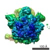 1417MC M: map data used to model this data C: citing same article ( |
|---|---|
| Similar structure data |


-Links
 PDBj
PDBj


- Assembly
| Deposited unit | 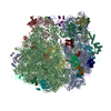
|
|---|---|
| 1 |
|
-Components
-RNA chain , 4 types, 4 molecules AABABBBC
| #1: RNA chain | Mass: 483490.531 Da / Num. of mol.: 1 / Source method: isolated from a natural source / Details: modeled using Escherichia coli 2AVY as template / Source: (natural) Spinacea oleracea (spinach) / Cellular location: chloroplast / References: GenBank: 7636084 |
|---|---|
| #22: RNA chain | Mass: 911368.312 Da / Num. of mol.: 1 / Source method: isolated from a natural source / Details: modeled using Escherichia coli 2AWB as template / Source: (natural) Spinacea oleracea (spinach) / References: EMBL: SOL400848 |
| #23: RNA chain | Mass: 37743.441 Da / Num. of mol.: 1 / Source method: isolated from a natural source / Details: modeled using Escherichia coli 2AWB as template / Source: (natural) Spinacea oleracea (spinach) / References: EMBL: SOL400848 |
| #24: RNA chain | Mass: 33330.867 Da / Num. of mol.: 1 / Source method: isolated from a natural source / Details: modeled using Escherichia coli 2AWB as template / Source: (natural) Spinacea oleracea (spinach) / References: EMBL: SOL400848 |
+Ribosomal Protein ... , 49 types, 49 molecules ABACADAEAFAGAHAIAJAKALAMANAOAPAQARASATAUBDBEBFBGBHBIBJBKBLBM...
-Experimental details
-Experiment
| Experiment | Method: ELECTRON MICROSCOPY |
|---|---|
| EM experiment | Aggregation state: PARTICLE / 3D reconstruction method: single particle reconstruction |
- Sample preparation
| Component | Name: spinach 70S chloro-ribosome / Type: RIBOSOME / Details: tight couple chloroplast 70S ribosomes |
|---|---|
| Buffer solution | Name: 10mM Tris-HCL pH 7.6, 50mM KCL, 10mM MgOAc, 7mM 2-ME / pH: 7.6 Details: 10mM Tris-HCL pH 7.6, 50mM KCL, 10mM MgOAc, 7mM 2-ME |
| Specimen | Embedding applied: NO / Shadowing applied: NO / Staining applied: NO / Vitrification applied: YES |
| Vitrification | Instrument: HOMEMADE PLUNGER / Cryogen name: ETHANE Details: 5 microliters applied to the grid then blotted for 3 seconds with Whatman number 1 filter paper before plunging in liquid ethane. |
- Electron microscopy imaging
| Experimental equipment |  Model: Tecnai F20 / Image courtesy: FEI Company |
|---|---|
| Microscopy | Model: FEI TECNAI F20 |
| Electron gun | Electron source:  FIELD EMISSION GUN / Accelerating voltage: 200 kV / Illumination mode: FLOOD BEAM FIELD EMISSION GUN / Accelerating voltage: 200 kV / Illumination mode: FLOOD BEAM |
| Electron lens | Mode: BRIGHT FIELD / Nominal magnification: 50000 X / Calibrated magnification: 50760 X / Nominal defocus max: 3500 nm / Nominal defocus min: 700 nm / Cs: 2 mm |
| Specimen holder | Temperature: 93 K / Tilt angle max: 0 ° / Tilt angle min: 0 ° |
| Image recording | Electron dose: 20 e/Å2 / Film or detector model: KODAK SO-163 FILM |
- Processing
| CTF correction | Details: CTF correction for each Micrograph | ||||||||||||
|---|---|---|---|---|---|---|---|---|---|---|---|---|---|
| Symmetry | Point symmetry: C1 (asymmetric) | ||||||||||||
| 3D reconstruction | Method: The projection matching procedure within the SPIDER software was used to get 3D map Resolution: 9.4 Å / Num. of particles: 86370 / Actual pixel size: 2.76 Å / Magnification calibration: 50,760 Details: An 11.5 A E.coli 70S ribosome map was used as initial reference and then resulting 18 A map from reconstruction of 70S chloro-ribosome was used as a reference for iterative refinement Symmetry type: POINT | ||||||||||||
| Atomic model building | Protocol: RIGID BODY FIT / Space: REAL / Target criteria: Best visual fit using the program O Details: METHOD--Cross-Correlation based manual fitting in O REFINEMENT PROTOCOL--Rigid Body | ||||||||||||
| Atomic model building | 3D fitting-ID: 1 / Details: 2XYZ AND 2ZXY FOR SMALL AND LARGE SUBUNIT RESPECTIVELY / Source name: PDB / Type: experimental model
| ||||||||||||
| Refinement step | Cycle: LAST
|