 Movie
Movie Controller

[English] 日本語
 Yorodumi
- EMDB-1417: Cryo-EM study of the Spinach chloroplast ribosome reveals the str... -
+ Open data

- Basic information
| Entry | Database: EMDB / ID: EMD-1417 | |||||||||
|---|---|---|---|---|---|---|---|---|---|---|
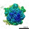
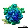
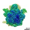
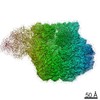
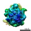
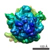
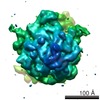
| Title | Cryo-EM study of the Spinach chloroplast ribosome reveals the structural and functional roles of plastid-specific ribosomal proteins | |||||||||
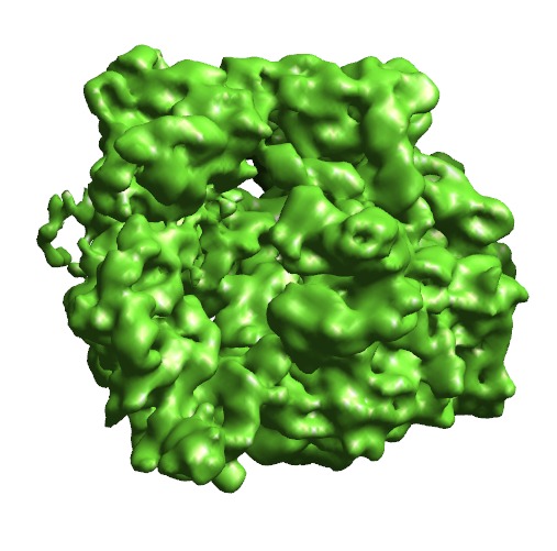
 Map data Map data | Spinach Chloroplast 70S ribosome | |||||||||
 Sample Sample |
| |||||||||
| Function / homology |  Function and homology information Function and homology informationplastid small ribosomal subunit / mitochondrial large ribosomal subunit / mitochondrial small ribosomal subunit / mitochondrial translation / chloroplast / DNA-templated transcription termination / large ribosomal subunit / transferase activity / ribosomal small subunit assembly / ribosome biogenesis ...plastid small ribosomal subunit / mitochondrial large ribosomal subunit / mitochondrial small ribosomal subunit / mitochondrial translation / chloroplast / DNA-templated transcription termination / large ribosomal subunit / transferase activity / ribosomal small subunit assembly / ribosome biogenesis / ribosomal small subunit biogenesis / ribosomal large subunit assembly / small ribosomal subunit / small ribosomal subunit rRNA binding / large ribosomal subunit rRNA binding / cytosolic small ribosomal subunit / cytosolic large ribosomal subunit / negative regulation of translation / rRNA binding / structural constituent of ribosome / ribosome / translation / ribonucleoprotein complex / response to antibiotic / mRNA binding / mitochondrion / RNA binding Similarity search - Function | |||||||||
| Biological species |  Spinacia oleracea (spinach) Spinacia oleracea (spinach) | |||||||||
| Method | single particle reconstruction / cryo EM / Resolution: 9.4 Å | |||||||||
 Authors Authors | Sharma MR / Wilson DN / Datta PP / Barat C / Schluenzen F / Fucini P / Agrawal RK | |||||||||
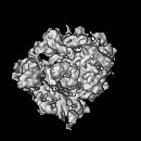
 Citation Citation | Journal: Proc Natl Acad Sci U S A / Year: 2007 Title: Cryo-EM study of the spinach chloroplast ribosome reveals the structural and functional roles of plastid-specific ribosomal proteins. Authors: Manjuli R Sharma / Daniel N Wilson / Partha P Datta / Chandana Barat / Frank Schluenzen / Paola Fucini / Rajendra K Agrawal /  Abstract: Protein synthesis in the chloroplast is carried out by chloroplast ribosomes (chloro-ribosome) and regulated in a light-dependent manner. Chloroplast or plastid ribosomal proteins (PRPs) generally ...Protein synthesis in the chloroplast is carried out by chloroplast ribosomes (chloro-ribosome) and regulated in a light-dependent manner. Chloroplast or plastid ribosomal proteins (PRPs) generally are larger than their bacterial counterparts, and chloro-ribosomes contain additional plastid-specific ribosomal proteins (PSRPs); however, it is unclear to what extent these proteins play structural or regulatory roles during translation. We have obtained a three-dimensional cryo-EM map of the spinach 70S chloro-ribosome, revealing the overall structural organization to be similar to bacterial ribosomes. Fitting of the conserved portions of the x-ray crystallographic structure of the bacterial 70S ribosome into our cryo-EM map of the chloro-ribosome reveals the positions of PRP extensions and the locations of the PSRPs. Surprisingly, PSRP1 binds in the decoding region of the small (30S) ribosomal subunit, in a manner that would preclude the binding of messenger and transfer RNAs to the ribosome, suggesting that PSRP1 is a translation factor rather than a ribosomal protein. PSRP2 and PSRP3 appear to structurally compensate for missing segments of the 16S rRNA within the 30S subunit, whereas PSRP4 occupies a position buried within the head of the 30S subunit. One of the two PSRPs in the large (50S) ribosomal subunit lies near the tRNA exit site. Furthermore, we find a mass of density corresponding to chloro-ribosome recycling factor; domain II of this factor appears to interact with the flexible C-terminal domain of PSRP1. Our study provides evolutionary insights into the structural and functional roles that the PSRPs play during protein synthesis in chloroplasts. | |||||||||
| History |
|
- Structure visualization
| Movie |
Movie viewer |
|---|---|
| Structure viewer | EM map: SurfViewMolmilJmol/JSmol |
| Supplemental images |
- Downloads & links
-EMDB archive
| Map data | emd_1417.map.gz | 7.6 MB | EMDB map data format | |
|---|---|---|---|---|
| Header (meta data) | emd-1417-v30.xmlemd-1417.xml | 10.3 KB 10.3 KB | Display Display | EMDB header |
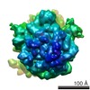
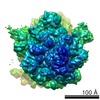
| Images |  1417-3BBN-3BBO.pngEMD-1417.tif 1417-3BBN-3BBO.pngEMD-1417.tif | 266 KB 744.4 KB | ||
| Archive directory |  http://ftp.pdbj.org/pub/emdb/structures/EMD-1417ftp://ftp.pdbj.org/pub/emdb/structures/EMD-1417 http://ftp.pdbj.org/pub/emdb/structures/EMD-1417ftp://ftp.pdbj.org/pub/emdb/structures/EMD-1417 | HTTPS FTP |
-Related structure data
| Related structure data |  4v61MC M: atomic model generated by this map C: citing same article ( |
|---|---|
| Similar structure data |

-Links
| EMDB pages | EMDB (EBI/PDBe) / EMDataResource |
|---|---|
| Related items in Molecule of the Month |


-Map
| File | Download / File: emd_1417.map.gz / Format: CCP4 / Size: 8.2 MB / Type: IMAGE STORED AS FLOATING POINT NUMBER (4 BYTES) | ||||||||||||||||||||||||||||||||||||||||||||||||||||||||||||||||||||
|---|---|---|---|---|---|---|---|---|---|---|---|---|---|---|---|---|---|---|---|---|---|---|---|---|---|---|---|---|---|---|---|---|---|---|---|---|---|---|---|---|---|---|---|---|---|---|---|---|---|---|---|---|---|---|---|---|---|---|---|---|---|---|---|---|---|---|---|---|---|
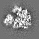
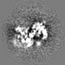
| Annotation | Spinach Chloroplast 70S ribosome | ||||||||||||||||||||||||||||||||||||||||||||||||||||||||||||||||||||
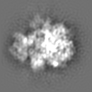
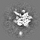
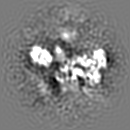
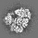
| Projections & slices | Image control
Images are generated by Spider. | ||||||||||||||||||||||||||||||||||||||||||||||||||||||||||||||||||||
| Voxel size | X=Y=Z: 2.76 Å | ||||||||||||||||||||||||||||||||||||||||||||||||||||||||||||||||||||
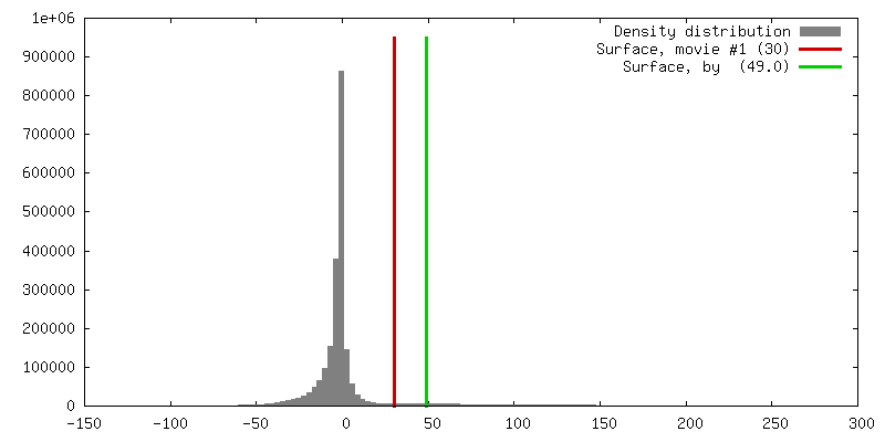
| Density |
| ||||||||||||||||||||||||||||||||||||||||||||||||||||||||||||||||||||
| Symmetry | Space group: 1 | ||||||||||||||||||||||||||||||||||||||||||||||||||||||||||||||||||||
| Details | EMDB XML:
CCP4 map header:
| ||||||||||||||||||||||||||||||||||||||||||||||||||||||||||||||||||||
 Z (Sec.)
 Y (Row.)
 X (Col.)


-Supplemental data
- Sample components
-Entire : Spinacea oleracea chloroplast 70S ribosome
| Entire | Name: Spinacea oleracea chloroplast 70S ribosome |
|---|---|
| Components |
|
-Supramolecule #1000: Spinacea oleracea chloroplast 70S ribosome
| Supramolecule | Name: Spinacea oleracea chloroplast 70S ribosome / type: sample / ID: 1000 / Number unique components: 1 |
|---|---|
| Molecular weight | Theoretical: 2.5 MDa |
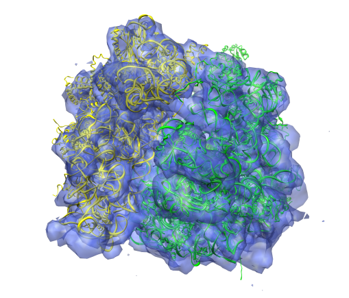
-Supramolecule #1: Spinach Chloroplast 70S Ribosome
| Supramolecule | Name: Spinach Chloroplast 70S Ribosome / type: complex / ID: 1 / Name.synonym: chloro-ribosome Details: The SSU 30S has PSRP1,2,3, and 4 identified. LSU 50S did not have PRPL25 and PRPL30 density present. pRRF (plastid ribosome recycling factor)is tightly bound to LSU 50S subunit. One of the ...Details: The SSU 30S has PSRP1,2,3, and 4 identified. LSU 50S did not have PRPL25 and PRPL30 density present. pRRF (plastid ribosome recycling factor)is tightly bound to LSU 50S subunit. One of the two PSRPs on the LSU 50S subunit is identified. Recombinant expression: No / Ribosome-details: ribosome-eukaryote: ALL |
|---|---|
| Source (natural) | Organism: Spinacia oleracea (spinach) / synonym: Spinach |
| Molecular weight | Experimental: 2.5 MDa |
-Experimental details
-Structure determination
| Method | cryo EM |
|---|---|
 Processing Processing | single particle reconstruction |
| Aggregation state | particle |
-Sample preparation
| Buffer | pH: 7.6 Details: 10mM Tris-HCL pH 7.6, 50mM KCL, 10mM MgOAc, 7mM 2-ME |
|---|---|
| Grid | Details: quantifoil 300 mesh copper grid |
| Vitrification | Cryogen name: ETHANE / Chamber humidity: 100 % / Chamber temperature: 277 K / Instrument: HOMEMADE PLUNGER / Details: Vitrification instrument: Cryo-plunger Method: 5 microliters applied to the grid then blotted for 3 seconds with Whatman number 1 filter paper before plunging |
- Electron microscopy
| Microscope | FEI TECNAI F20 |
|---|---|
| Temperature | Average: 93 K |
| Alignment procedure | Legacy - Astigmatism: objective lens astigmatism was corrected at 250K times magnification |
| Image recording | Category: FILM / Film or detector model: KODAK SO-163 FILM / Digitization - Scanner: ZEISS SCAI / Digitization - Sampling interval: 14 µm / Number real images: 164 / Average electron dose: 20 e/Å2 / Bits/pixel: 12 |
| Electron beam | Acceleration voltage: 200 kV / Electron source:  FIELD EMISSION GUN FIELD EMISSION GUN |
| Electron optics | Calibrated magnification: 50760 / Illumination mode: FLOOD BEAM / Imaging mode: BRIGHT FIELD / Nominal defocus max: 4.4 µm / Nominal defocus min: 1.4 µm / Nominal magnification: 50000 |
| Sample stage | Specimen holder: Cryo Transfer Holder / Specimen holder model: OTHER |
| Experimental equipment |  Model: Tecnai F20 / Image courtesy: FEI Company |
-Image processing
| Details | Initially, 192,133 images were selected using automated particle picking program |
|---|---|
| CTF correction | Details: each micrograph |
| Final reconstruction | Applied symmetry - Point group: C1 (asymmetric) / Algorithm: OTHER / Resolution.type: BY AUTHOR / Resolution: 9.4 Å / Resolution method: FSC 0.5 CUT-OFF / Software - Name: SPIDER / Number images used: 86370 |
| Final two d classification | Number classes: 83 |
-Atomic model buiding 1
| Initial model | PDB ID: |
|---|---|
| Software | Name: O |
| Details | Protocol: Rigid Body. Docking of crystallographic structures in 3D map was performed using program O |
| Refinement | Protocol: RIGID BODY FIT |
| Output model | PDB-4v61: |

