[English] 日本語
Yorodumi
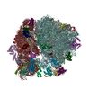
- PDB-4v61: Homology model for the Spinach chloroplast 30S subunit fitted to ... -

+
Open data


ID or keywords:

Loading...

-
Basic information

Entry
Database: PDB / ID: 4v61
TitleHomology model for the Spinach chloroplast 30S subunit fitted to 9.4A cryo-EM map of the 70S chlororibosome.
Components
  • (Ribosomal Protein ...) x 49
  • 16S rRNA
  • 23S rRNA
  • 4.8S rRNA
  • 5S rRNA
KeywordsRIBOSOME / SMALL RIBOSOMAL SUBUNIT / SPINACH CHLOROPLAST RIBOSOME / RIBONUCLEOPROTEIN PARTICLE / MACROMOLECULAR COMPLEX
Function / homology
Function and homology information


plastid small ribosomal subunit / mitochondrial large ribosomal subunit / mitochondrial small ribosomal subunit / mitochondrial translation / chloroplast / DNA-templated transcription termination / ribosomal small subunit biogenesis / small ribosomal subunit rRNA binding / large ribosomal subunit / ribosomal small subunit assembly ...plastid small ribosomal subunit / mitochondrial large ribosomal subunit / mitochondrial small ribosomal subunit / mitochondrial translation / chloroplast / DNA-templated transcription termination / ribosomal small subunit biogenesis / small ribosomal subunit rRNA binding / large ribosomal subunit / ribosomal small subunit assembly / small ribosomal subunit / large ribosomal subunit rRNA binding / transferase activity / cytosolic small ribosomal subunit / ribosomal large subunit assembly / cytoplasmic translation / cytosolic large ribosomal subunit / negative regulation of translation / rRNA binding / ribosome / structural constituent of ribosome / ribonucleoprotein complex / translation / response to antibiotic / mRNA binding / mitochondrion / RNA binding
Similarity search - Function
Ribosomal protein L32p, plant/cyanobacteria type / Ribosomal protein L1, bacterial-type / Ribosomal protein L1, conserved site / Ribosomal protein L1 signature. / Ribosomal protein L1, 3-layer alpha/beta-sandwich / Ribosomal protein S14, bacterial/plastid / Ribosomal protein L1-like / Ribosomal protein L1/ribosomal biogenesis protein / Ribosomal protein L1p/L10e family / Ribosomal protein L11, bacterial-type ...Ribosomal protein L32p, plant/cyanobacteria type / Ribosomal protein L1, bacterial-type / Ribosomal protein L1, conserved site / Ribosomal protein L1 signature. / Ribosomal protein L1, 3-layer alpha/beta-sandwich / Ribosomal protein S14, bacterial/plastid / Ribosomal protein L1-like / Ribosomal protein L1/ribosomal biogenesis protein / Ribosomal protein L1p/L10e family / Ribosomal protein L11, bacterial-type / Ribosomal protein S16, conserved site / Ribosomal protein S16 signature. / Ribosomal protein L21, conserved site / Ribosomal protein L21 signature. / Ribosomal protein L11, conserved site / Ribosomal protein L11 signature. / Ribosomal protein L16 signature 1. / Ribosomal protein L16, conserved site / Ribosomal protein L16 signature 2. / Ribosomal protein L11, N-terminal / Ribosomal protein L11, N-terminal domain / Ribosomal protein L11/L12 / Ribosomal protein L11, C-terminal / Ribosomal protein L11, C-terminal domain superfamily / Ribosomal protein L11/L12, N-terminal domain superfamily / Ribosomal protein L11, RNA binding domain / Ribosomal protein L11/L12 / : / Ribosomal protein L35, conserved site / Ribosomal protein L35 signature. / Ribosomal protein L33, conserved site / Ribosomal protein L33 signature. / Ribosomal protein L35, non-mitochondrial / Ribosomal protein L5, bacterial-type / Ribosomal protein S3, bacterial-type / Ribosomal protein S19, bacterial-type / Ribosomal protein S7, bacterial/organellar-type / Ribosomal protein S11, bacterial-type / Ribosomal protein L20 signature. / Ribosomal protein S9, bacterial/plastid / Ribosomal protein S4, bacterial-type / Ribosomal protein S5, bacterial-type / Ribosomal protein L14P, bacterial-type / Ribosomal protein S6, plastid/chloroplast / Ribosomal protein L22, bacterial/chloroplast-type / Ribosomal protein L2, bacterial/organellar-type / Ribosomal protein S2, bacteria/mitochondria/plastid / Ribosomal protein L35 / Ribosomal protein L35 superfamily / Ribosomal protein L35 / Ribosomal protein L33 / Ribosomal protein L33 / Ribosomal protein L33 superfamily / Ribosomal protein S18, conserved site / Ribosomal protein S18 signature. / Ribosomal protein L16 / Ribosomal protein S16 / Ribosomal protein S16 domain superfamily / Ribosomal protein S16 / : / Ribosomal protein L20 / Ribosomal protein S15, bacterial-type / Ribosomal protein L20 / Ribosomal protein L20, C-terminal / Ribosomal protein L21 / Ribosomal protein L19 / Ribosomal protein L19 / Ribosomal protein S6 / Ribosomal protein S6 / Ribosomal protein L19 superfamily / Ribosomal proteins 50S L24/mitochondrial 39S L24 / Ribosomal protein S6 superfamily / Ribosomal protein L21-like / L21-like superfamily / Ribosomal prokaryotic L21 protein / Ribosomal protein S12, bacterial-type / Ribosomal protein L32p / Ribosomal protein L24 / Translation elongation factor EF1B/ribosomal protein S6 / Ribosomal protein L13, bacterial-type / Ribosomal protein S2 signature 2. / Ribosomal protein S18 / Ribosomal protein S18 / Ribosomal protein S18 superfamily / 50S ribosomal protein uL4 / Ribosomal protein L34 / Ribosomal protein L34 / Ribosomal protein S3, conserved site / Ribosomal protein S3 signature. / Ribosomal protein S14, conserved site / Ribosomal protein S14 signature. / Ribosomal protein L5, conserved site / Ribosomal protein L5 signature. / Ribosomal protein S2 signature 1. / Ribosomal protein L2 signature. / Type-2 KH domain profile. / K Homology domain, type 2 / Ribosomal protein S3, C-terminal / Ribosomal protein S3, C-terminal domain / Ribosomal protein S3, C-terminal domain superfamily
Similarity search - Domain/homology
: / : / RNA / RNA (> 10) / RNA (> 100) / RNA (> 1000) / Large ribosomal subunit protein uL4c / Small ribosomal subunit protein uS11c / Small ribosomal subunit protein uS14c / Small ribosomal subunit protein uS19c ...: / : / RNA / RNA (> 10) / RNA (> 100) / RNA (> 1000) / Large ribosomal subunit protein uL4c / Small ribosomal subunit protein uS11c / Small ribosomal subunit protein uS14c / Small ribosomal subunit protein uS19c / Large ribosomal subunit protein uL2cz/uL2cy / Small ribosomal subunit protein uS2c / Large ribosomal subunit protein uL22c / Small ribosomal subunit protein uS3c / Large ribosomal subunit protein uL14c / Small ribosomal subunit protein uS8c / Large ribosomal subunit protein uL13c / Small ribosomal subunit protein uS4c / Large ribosomal subunit protein uL16c / Large ribosomal subunit protein bL35c / Large ribosomal subunit protein bL21c / Large ribosomal subunit protein uL24c / Large ribosomal subunit protein bL20c / Large ribosomal subunit protein bL32c / Large ribosomal subunit protein bL33c / Small ribosomal subunit protein bS16c / Large ribosomal subunit protein uL11c / Small ribosomal subunit protein uS12cz/uS12cy / Small ribosomal subunit protein uS7cz/uS7cy / Small ribosomal subunit protein uS13c / Large ribosomal subunit protein uL5c / Large ribosomal subunit protein bL34c / Small ribosomal subunit protein uS9c / Small ribosomal subunit protein bS6c alpha / Large ribosomal subunit protein bL19c / Large ribosomal subunit protein uL1c / Large ribosomal subunit protein uL23c / Small ribosomal subunit protein uS15c / Small ribosomal subunit protein bS18c / Small ribosomal subunit protein uS5c
Similarity search - Component
Biological speciesSpinacea oleracea (spinach)
MethodELECTRON MICROSCOPY / single particle reconstruction / cryo EM / Resolution: 9.4 Å
AuthorsSharma, M.R. / Wilson, D.N. / Datta, P.P. / Barat, C. / Schluenzen, F. / Fucini, P. / Agrawal, R.K.
CitationJournal: Proc Natl Acad Sci U S A / Year: 2007
Title: Cryo-EM study of the spinach chloroplast ribosome reveals the structural and functional roles of plastid-specific ribosomal proteins.
Authors: Manjuli R Sharma / Daniel N Wilson / Partha P Datta / Chandana Barat / Frank Schluenzen / Paola Fucini / Rajendra K Agrawal /
Abstract: Protein synthesis in the chloroplast is carried out by chloroplast ribosomes (chloro-ribosome) and regulated in a light-dependent manner. Chloroplast or plastid ribosomal proteins (PRPs) generally ...Protein synthesis in the chloroplast is carried out by chloroplast ribosomes (chloro-ribosome) and regulated in a light-dependent manner. Chloroplast or plastid ribosomal proteins (PRPs) generally are larger than their bacterial counterparts, and chloro-ribosomes contain additional plastid-specific ribosomal proteins (PSRPs); however, it is unclear to what extent these proteins play structural or regulatory roles during translation. We have obtained a three-dimensional cryo-EM map of the spinach 70S chloro-ribosome, revealing the overall structural organization to be similar to bacterial ribosomes. Fitting of the conserved portions of the x-ray crystallographic structure of the bacterial 70S ribosome into our cryo-EM map of the chloro-ribosome reveals the positions of PRP extensions and the locations of the PSRPs. Surprisingly, PSRP1 binds in the decoding region of the small (30S) ribosomal subunit, in a manner that would preclude the binding of messenger and transfer RNAs to the ribosome, suggesting that PSRP1 is a translation factor rather than a ribosomal protein. PSRP2 and PSRP3 appear to structurally compensate for missing segments of the 16S rRNA within the 30S subunit, whereas PSRP4 occupies a position buried within the head of the 30S subunit. One of the two PSRPs in the large (50S) ribosomal subunit lies near the tRNA exit site. Furthermore, we find a mass of density corresponding to chloro-ribosome recycling factor; domain II of this factor appears to interact with the flexible C-terminal domain of PSRP1. Our study provides evolutionary insights into the structural and functional roles that the PSRPs play during protein synthesis in chloroplasts.
History
DepositionNov 9, 2007Deposition site: RCSB / Processing site: RCSB
Revision 1.0Jul 9, 2014Provider: repository / Type: Initial release
SupersessionDec 10, 2014ID: 3BBO, 3BBN
Revision 1.1Dec 10, 2014Group: Other
Revision 1.2Mar 25, 2015Group: Other
Revision 1.3Dec 18, 2019Group: Data collection / Database references / Other
Category: atom_sites / em_image_scans ...atom_sites / em_image_scans / struct_ref_seq / struct_ref_seq_dif
Item: _atom_sites.fract_transf_matrix[1][1] / _atom_sites.fract_transf_matrix[2][2] ..._atom_sites.fract_transf_matrix[1][1] / _atom_sites.fract_transf_matrix[2][2] / _atom_sites.fract_transf_matrix[3][3] / _struct_ref_seq.db_align_beg / _struct_ref_seq.db_align_end / _struct_ref_seq_dif.details
Revision 1.4Feb 28, 2024Group: Data collection / Database references / Refinement description
Category: chem_comp_atom / chem_comp_bond ...chem_comp_atom / chem_comp_bond / database_2 / em_3d_fitting_list / pdbx_initial_refinement_model
Item: _database_2.pdbx_DOI / _database_2.pdbx_database_accession ..._database_2.pdbx_DOI / _database_2.pdbx_database_accession / _em_3d_fitting_list.accession_code / _em_3d_fitting_list.initial_refinement_model_id / _em_3d_fitting_list.source_name / _em_3d_fitting_list.type

-
Structure visualization

Movie
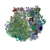
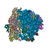
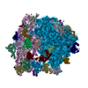
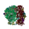
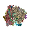
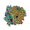
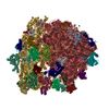
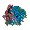
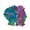
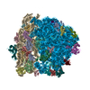
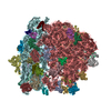
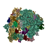
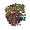
  • Deposited structure unit
  • Imaged by Jmol
  • Download
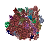
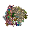
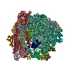
  • Superimposition on EM map
  • EMDB-1417
  • Imaged by UCSF Chimera
  • Download
Movie viewer
Structure viewerMolecule:
MolmilJmol/JSmol

Downloads & links

-
Assembly

Deposited unit
AA: 16S rRNA
AB: Ribosomal Protein S2
AC: Ribosomal Protein S3
AD: Ribosomal Protein S4
AE: Ribosomal Protein S5
AF: Ribosomal Protein S6
AG: Ribosomal Protein S7
AH: Ribosomal Protein S8
AI: Ribosomal Protein S9
AJ: Ribosomal Protein S10
AK: Ribosomal Protein S11
AL: Ribosomal Protein S12
AM: Ribosomal Protein S13
AN: Ribosomal Protein S14
AO: Ribosomal Protein S15
AP: Ribosomal Protein S16
AQ: Ribosomal Protein S17
AR: Ribosomal Protein S18
AS: Ribosomal Protein S19
AT: Ribosomal Protein S20
AU: Ribosomal Protein S21
BA: 23S rRNA
BB: 5S rRNA
BC: 4.8S rRNA
BD: Ribosomal Protein L1
BE: Ribosomal Protein L2
BF: Ribosomal Protein L3
BG: Ribosomal Protein L4
BH: Ribosomal Protein L5
BI: Ribosomal Protein L6
BJ: Ribosomal Protein L9
BK: Ribosomal Protein L11
BL: Ribosomal Protein L13
BM: Ribosomal Protein L14
BN: Ribosomal Protein L15
BO: Ribosomal Protein L16
BP: Ribosomal Protein L17
BQ: Ribosomal Protein L18
BR: Ribosomal Protein L19
BS: Ribosomal Protein L20
BT: Ribosomal Protein L21
BU: Ribosomal Protein L22
BV: Ribosomal Protein L23
BW: Ribosomal Protein L24
BX: Ribosomal Protein L27
BY: Ribosomal Protein L28
BZ: Ribosomal Protein L29
B1: Ribosomal Protein L31
B2: Ribosomal Protein L32
B3: Ribosomal Protein L33
B4: Ribosomal Protein L34
B5: Ribosomal Protein L35
B6: Ribosomal Protein L36


Theoretical massNumber of molelcules
Total (without water)2,447,89153
Polymers2,447,89153
Non-polymers00
Water00
1


  • Idetical with deposited unit
  • defined by author&software
TypeNameSymmetry operationNumber
identity operation1_555x,y,z1
MethodPISA

-
Components

-
RNA chain , 4 types, 4 molecules AABABBBC

#1: RNA chain 16S rRNA


Mass: 483490.531 Da / Num. of mol.: 1 / Source method: isolated from a natural source / Details: modeled using Escherichia coli 2AVY as template / Source: (natural) Spinacea oleracea (spinach) / Cellular location: chloroplast / References: GenBank: 7636084
#22: RNA chain 23S rRNA


Mass: 911368.312 Da / Num. of mol.: 1 / Source method: isolated from a natural source / Details: modeled using Escherichia coli 2AWB as template / Source: (natural) Spinacea oleracea (spinach) / References: EMBL: SOL400848
#23: RNA chain 5S rRNA


Mass: 37743.441 Da / Num. of mol.: 1 / Source method: isolated from a natural source / Details: modeled using Escherichia coli 2AWB as template / Source: (natural) Spinacea oleracea (spinach) / References: EMBL: SOL400848
#24: RNA chain 4.8S rRNA


Mass: 33330.867 Da / Num. of mol.: 1 / Source method: isolated from a natural source / Details: modeled using Escherichia coli 2AWB as template / Source: (natural) Spinacea oleracea (spinach) / References: EMBL: SOL400848

+
Ribosomal Protein ... , 49 types, 49 molecules ABACADAEAFAGAHAIAJAKALAMANAOAPAQARASATAUBDBEBFBGBHBIBJBKBLBM...

#2: Protein Ribosomal Protein S2


Mass: 26092.336 Da / Num. of mol.: 1 / Source method: isolated from a natural source / Details: modeled using Escherichia coli 2AVY as template / Source: (natural) Spinacea oleracea (spinach) / Cellular location: chloroplast / References: UniProt: P08242
#3: Protein Ribosomal Protein S3


Mass: 24965.156 Da / Num. of mol.: 1 / Source method: isolated from a natural source / Details: modeled using Escherichia coli 2AVY as template / Source: (natural) Spinacea oleracea (spinach) / Cellular location: chloroplast / References: UniProt: P09595
#4: Protein Ribosomal Protein S4


Mass: 23454.531 Da / Num. of mol.: 1 / Source method: isolated from a natural source / Details: modeled using Escherichia coli 2AVY as template / Source: (natural) Spinacea oleracea (spinach) / Cellular location: chloroplast / References: UniProt: P13788
#5: Protein Ribosomal Protein S5


Mass: 33626.363 Da / Num. of mol.: 1 / Source method: isolated from a natural source / Details: modeled using Escherichia coli 2AVY as template / Source: (natural) Spinacea oleracea (spinach) / Cellular location: chloroplast / References: UniProt: Q9ST69
#6: Protein Ribosomal Protein S6


Mass: 18870.361 Da / Num. of mol.: 1 / Source method: isolated from a natural source / Details: modeled using Escherichia coli 2AVY as template / Source: (natural) Spinacea oleracea (spinach) / Cellular location: chloroplast / References: UniProt: P82403
#7: Protein Ribosomal Protein S7


Mass: 17378.309 Da / Num. of mol.: 1 / Source method: isolated from a natural source / Details: modeled using Escherichia coli 2AVY as template / Source: (natural) Spinacea oleracea (spinach) / Cellular location: chloroplast / References: UniProt: P82129
#8: Protein Ribosomal Protein S8


Mass: 15527.256 Da / Num. of mol.: 1 / Source method: isolated from a natural source / Details: modeled using Escherichia coli 2AVY as template / Source: (natural) Spinacea oleracea (spinach) / Cellular location: chloroplast / References: UniProt: P09597
#9: Protein Ribosomal Protein S9


Mass: 21350.842 Da / Num. of mol.: 1 / Source method: isolated from a natural source / Details: modeled using Escherichia coli 2AVY as template / Source: (natural) Spinacea oleracea (spinach) / Cellular location: chloroplast / References: UniProt: P82278
#10: Protein Ribosomal Protein S10


Mass: 21632.629 Da / Num. of mol.: 1 / Source method: isolated from a natural source / Details: modeled using Escherichia coli 2AVY as template / Source: (natural) Spinacea oleracea (spinach) / Cellular location: chloroplast
#11: Protein Ribosomal Protein S11


Mass: 15085.706 Da / Num. of mol.: 1 / Source method: isolated from a natural source / Details: modeled using Escherichia coli 2AVY as template / Source: (natural) Spinacea oleracea (spinach) / Cellular location: chloroplast / References: UniProt: P06506
#12: Protein Ribosomal Protein S12


Mass: 13794.261 Da / Num. of mol.: 1 / Source method: isolated from a natural source / Details: modeled using Escherichia coli 2AVY as template / Source: (natural) Spinacea oleracea (spinach) / Cellular location: chloroplast / References: UniProt: P62128
#13: Protein Ribosomal Protein S13


Mass: 16306.087 Da / Num. of mol.: 1 / Source method: isolated from a natural source / Details: modeled using Escherichia coli 2AVY as template / Source: (natural) Spinacea oleracea (spinach) / Cellular location: chloroplast / References: UniProt: P82163
#14: Protein Ribosomal Protein S14


Mass: 11809.832 Da / Num. of mol.: 1 / Source method: isolated from a natural source / Details: modeled using Escherichia coli 2AVY as template / Source: (natural) Spinacea oleracea (spinach) / Cellular location: chloroplast / References: UniProt: P06507
#15: Protein Ribosomal Protein S15


Mass: 10778.763 Da / Num. of mol.: 1 / Source method: isolated from a natural source / Details: modeled using Escherichia coli 2AVY as template / Source: (natural) Spinacea oleracea (spinach) / Cellular location: chloroplast / References: UniProt: Q9M3I4
#16: Protein Ribosomal Protein S16


Mass: 10454.237 Da / Num. of mol.: 1 / Source method: isolated from a natural source / Details: modeled using Escherichia coli 2AVY as template / Source: (natural) Spinacea oleracea (spinach) / Cellular location: chloroplast / References: UniProt: P28807
#17: Protein Ribosomal Protein S17


Mass: 15828.694 Da / Num. of mol.: 1 / Source method: isolated from a natural source / Details: modeled using Escherichia coli 2AVY as template / Source: (natural) Spinacea oleracea (spinach) / Cellular location: chloroplast
#18: Protein Ribosomal Protein S18


Mass: 12337.430 Da / Num. of mol.: 1 / Source method: isolated from a natural source / Details: modeled using Escherichia coli 2AVY as template / Source: (natural) Spinacea oleracea (spinach) / Cellular location: chloroplast / References: UniProt: Q9M3K7
#19: Protein Ribosomal Protein S19


Mass: 10632.500 Da / Num. of mol.: 1 / Source method: isolated from a natural source / Details: modeled using Escherichia coli 2AVY as template / Source: (natural) Spinacea oleracea (spinach) / Cellular location: chloroplast / References: UniProt: P06508
#20: Protein Ribosomal Protein S20


Mass: 21866.373 Da / Num. of mol.: 1 / Source method: isolated from a natural source / Details: modeled using Escherichia coli 2AVY as template / Source: (natural) Spinacea oleracea (spinach) / Cellular location: chloroplast
#21: Protein Ribosomal Protein S21


Mass: 21701.236 Da / Num. of mol.: 1 / Source method: isolated from a natural source / Details: modeled using Escherichia coli 2AVY as template / Source: (natural) Spinacea oleracea (spinach) / Cellular location: chloroplast
#25: Protein Ribosomal Protein L1


Mass: 38681.484 Da / Num. of mol.: 1 / Source method: isolated from a natural source / Details: modeled using Escherichia coli 2AWB as template / Source: (natural) Spinacea oleracea (spinach) / References: UniProt: Q9LE95
#26: Protein Ribosomal Protein L2


Mass: 29480.168 Da / Num. of mol.: 1 / Source method: isolated from a natural source / Details: modeled using Escherichia coli 2AWB as template / Source: (natural) Spinacea oleracea (spinach) / References: UniProt: P06509
#27: Protein Ribosomal Protein L3


Mass: 28423.457 Da / Num. of mol.: 1 / Source method: isolated from a natural source / Details: modeled using Escherichia coli 2AWB as template / Source: (natural) Spinacea oleracea (spinach)
#28: Protein Ribosomal Protein L4


Mass: 32479.617 Da / Num. of mol.: 1 / Source method: isolated from a natural source / Details: modeled using Escherichia coli 2AWB as template / Source: (natural) Spinacea oleracea (spinach) / References: UniProt: O49937
#29: Protein Ribosomal Protein L5


Mass: 24248.189 Da / Num. of mol.: 1 / Source method: isolated from a natural source / Details: modeled using Escherichia coli 2AWB as template / Source: (natural) Spinacea oleracea (spinach) / References: UniProt: P82192
#30: Protein Ribosomal Protein L6


Mass: 24747.686 Da / Num. of mol.: 1 / Source method: isolated from a natural source / Details: modeled using Escherichia coli 2AWB as template / Source: (natural) Spinacea oleracea (spinach)
#31: Protein Ribosomal Protein L9


Mass: 22169.826 Da / Num. of mol.: 1 / Source method: isolated from a natural source / Details: modeled using Escherichia coli 2AWB as template / Source: (natural) Spinacea oleracea (spinach)
#32: Protein Ribosomal Protein L11


Mass: 23689.980 Da / Num. of mol.: 1 / Source method: isolated from a natural source / Details: modeled using Escherichia coli 2AWB as template / Source: (natural) Spinacea oleracea (spinach) / References: UniProt: P31164
#33: Protein Ribosomal Protein L13


Mass: 28211.629 Da / Num. of mol.: 1 / Source method: isolated from a natural source / Details: modeled using Escherichia coli 2AWB as template / Source: (natural) Spinacea oleracea (spinach) / References: UniProt: P12629
#34: Protein Ribosomal Protein L14


Mass: 13484.741 Da / Num. of mol.: 1 / Source method: isolated from a natural source / Details: modeled using Escherichia coli 2AWB as template / Source: (natural) Spinacea oleracea (spinach) / References: UniProt: P09596
#35: Protein Ribosomal Protein L15


Mass: 27491.221 Da / Num. of mol.: 1 / Source method: isolated from a natural source / Details: modeled using Escherichia coli 2AWB as template / Source: (natural) Spinacea oleracea (spinach)
#36: Protein Ribosomal Protein L16


Mass: 15328.068 Da / Num. of mol.: 1 / Source method: isolated from a natural source / Details: modeled using Escherichia coli 2AWB as template / Source: (natural) Spinacea oleracea (spinach) / References: UniProt: P17353
#37: Protein Ribosomal Protein L17


Mass: 22983.852 Da / Num. of mol.: 1 / Source method: isolated from a natural source / Details: modeled using Escherichia coli 2AWB as template / Source: (natural) Spinacea oleracea (spinach)
#38: Protein Ribosomal Protein L18


Mass: 17850.727 Da / Num. of mol.: 1 / Source method: isolated from a natural source / Details: modeled using Escherichia coli 2AWB as template / Source: (natural) Spinacea oleracea (spinach)
#39: Protein Ribosomal Protein L19


Mass: 26121.254 Da / Num. of mol.: 1 / Source method: isolated from a natural source / Details: modeled using Escherichia coli 2AWB as template / Source: (natural) Spinacea oleracea (spinach) / References: UniProt: P82413
#40: Protein Ribosomal Protein L20


Mass: 14617.331 Da / Num. of mol.: 1 / Source method: isolated from a natural source / Details: modeled using Escherichia coli 2AWB as template / Source: (natural) Spinacea oleracea (spinach) / References: UniProt: P28803
#41: Protein Ribosomal Protein L21


Mass: 28554.883 Da / Num. of mol.: 1 / Source method: isolated from a natural source / Details: modeled using Escherichia coli 2AWB as template / Source: (natural) Spinacea oleracea (spinach) / References: UniProt: P24613
#42: Protein Ribosomal Protein L22


Mass: 23276.654 Da / Num. of mol.: 1 / Source method: isolated from a natural source / Details: modeled using Escherichia coli 2AWB as template / Source: (natural) Spinacea oleracea (spinach) / References: UniProt: P09594
#43: Protein Ribosomal Protein L23


Mass: 21832.164 Da / Num. of mol.: 1 / Source method: isolated from a natural source / Details: modeled using Escherichia coli 2AWB as template / Source: (natural) Spinacea oleracea (spinach) / References: UniProt: Q9LWB5
#44: Protein Ribosomal Protein L24


Mass: 21481.088 Da / Num. of mol.: 1 / Source method: isolated from a natural source / Details: modeled using Escherichia coli 2AWB as template / Source: (natural) Spinacea oleracea (spinach) / References: UniProt: P27683
#45: Protein Ribosomal Protein L27


Mass: 21771.951 Da / Num. of mol.: 1 / Source method: isolated from a natural source / Details: modeled using Escherichia coli 2AWB as template / Source: (natural) Spinacea oleracea (spinach)
#46: Protein Ribosomal Protein L28


Mass: 16730.691 Da / Num. of mol.: 1 / Source method: isolated from a natural source
Details: modeled using Thermus thermophilus 2J01 as template
Source: (natural) Spinacea oleracea (spinach)
#47: Protein Ribosomal Protein L29


Mass: 19415.543 Da / Num. of mol.: 1 / Source method: isolated from a natural source / Details: modeled using Escherichia coli 2AWB as template / Source: (natural) Spinacea oleracea (spinach)
#48: Protein Ribosomal Protein L31


Mass: 16060.906 Da / Num. of mol.: 1 / Source method: isolated from a natural source
Details: modeled using Thermus thermophilus 2J01 as template
Source: (natural) Spinacea oleracea (spinach)
#49: Protein Ribosomal Protein L32


Mass: 6650.969 Da / Num. of mol.: 1 / Source method: isolated from a natural source / Details: modeled using Escherichia coli 2AWB as template / Source: (natural) Spinacea oleracea (spinach) / References: UniProt: P28804
#50: Protein Ribosomal Protein L33


Mass: 7668.121 Da / Num. of mol.: 1 / Source method: isolated from a natural source / Details: modeled using Escherichia coli 2AWB as template / Source: (natural) Spinacea oleracea (spinach) / References: UniProt: P28805
#51: Protein Ribosomal Protein L34


Mass: 16126.633 Da / Num. of mol.: 1 / Source method: isolated from a natural source / Details: modeled using Escherichia coli 2AWB as template / Source: (natural) Spinacea oleracea (spinach) / References: UniProt: P82244
#52: Protein Ribosomal Protein L35


Mass: 17376.322 Da / Num. of mol.: 1 / Source method: isolated from a natural source / Details: modeled using Escherichia coli 2AWB as template / Source: (natural) Spinacea oleracea (spinach) / References: UniProt: P23326
#53: Protein Ribosomal Protein L36


Mass: 11509.490 Da / Num. of mol.: 1 / Source method: isolated from a natural source / Details: modeled using Escherichia coli 2AWB as template / Source: (natural) Spinacea oleracea (spinach)

-
Experimental details

-
Experiment

ExperimentMethod: ELECTRON MICROSCOPY
EM experimentAggregation state: PARTICLE / 3D reconstruction method: single particle reconstruction

-
Sample preparation

ComponentName: spinach 70S chloro-ribosome / Type: RIBOSOME / Details: tight couple chloroplast 70S ribosomes
Buffer solutionName: 10mM Tris-HCL pH 7.6, 50mM KCL, 10mM MgOAc, 7mM 2-ME / pH: 7.6
Details: 10mM Tris-HCL pH 7.6, 50mM KCL, 10mM MgOAc, 7mM 2-ME
SpecimenEmbedding applied: NO / Shadowing applied: NO / Staining applied: NO / Vitrification applied: YES
VitrificationInstrument: HOMEMADE PLUNGER / Cryogen name: ETHANE
Details: 5 microliters applied to the grid then blotted for 3 seconds with Whatman number 1 filter paper before plunging in liquid ethane.

-
Electron microscopy imaging

Experimental equipment
Model: Tecnai F20 / Image courtesy: FEI Company
MicroscopyModel: FEI TECNAI F20
Electron gunElectron source: FIELD EMISSION GUN / Accelerating voltage: 200 kV / Illumination mode: FLOOD BEAM
Electron lensMode: BRIGHT FIELD / Nominal magnification: 50000 X / Calibrated magnification: 50760 X / Nominal defocus max: 3500 nm / Nominal defocus min: 700 nm / Cs: 2 mm
Specimen holderTemperature: 93 K / Tilt angle max: 0 ° / Tilt angle min: 0 °
Image recordingElectron dose: 20 e/Å2 / Film or detector model: KODAK SO-163 FILM

-
Processing

CTF correctionDetails: CTF correction for each Micrograph
SymmetryPoint symmetry: C1 (asymmetric)
3D reconstructionMethod: The projection matching procedure within the SPIDER software was used to get 3D map
Resolution: 9.4 Å / Num. of particles: 86370 / Actual pixel size: 2.76 Å / Magnification calibration: 50,760
Details: An 11.5 A E.coli 70S ribosome map was used as initial reference and then resulting 18 A map from reconstruction of 70S chloro-ribosome was used as a reference for iterative refinement
Symmetry type: POINT
Atomic model buildingProtocol: RIGID BODY FIT / Space: REAL / Target criteria: Best visual fit using the program O
Details: METHOD--Cross-Correlation based manual fitting in O REFINEMENT PROTOCOL--Rigid Body
Atomic model building

3D fitting-ID: 1 / Details: 2XYZ AND 2ZXY FOR SMALL AND LARGE SUBUNIT RESPECTIVELY / Source name: PDB / Type: experimental model

IDPDB-IDAccession codeInitial refinement model-ID
12XYZ2XYZ1
22ZXY2ZXY2
Refinement stepCycle: LAST
ProteinNucleic acidLigandSolventTotal
Num. atoms19373 31745 0 0 51118

+
About Yorodumi

-
News

-
Feb 9, 2022. New format data for meta-information of EMDB entries

New format data for meta-information of EMDB entries

  • Version 3 of the EMDB header file is now the official format.
  • The previous official version 1.9 will be removed from the archive.

Related info.:EMDB header

External links:wwPDB to switch to version 3 of the EMDB data model

-
Aug 12, 2020. Covid-19 info

Covid-19 info

URL: https://pdbj.org/emnavi/covid19.php

New page: Covid-19 featured information page in EM Navigator.

Related info.:Covid-19 info / Mar 5, 2020. Novel coronavirus structure data

+
Mar 5, 2020. Novel coronavirus structure data

Novel coronavirus structure data

Related info.:Yorodumi Speices / Aug 12, 2020. Covid-19 info

External links:COVID-19 featured content - PDBj / Molecule of the Month (242):Coronavirus Proteases

+
Jan 31, 2019. EMDB accession codes are about to change! (news from PDBe EMDB page)

EMDB accession codes are about to change! (news from PDBe EMDB page)

  • The allocation of 4 digits for EMDB accession codes will soon come to an end. Whilst these codes will remain in use, new EMDB accession codes will include an additional digit and will expand incrementally as the available range of codes is exhausted. The current 4-digit format prefixed with “EMD-” (i.e. EMD-XXXX) will advance to a 5-digit format (i.e. EMD-XXXXX), and so on. It is currently estimated that the 4-digit codes will be depleted around Spring 2019, at which point the 5-digit format will come into force.
  • The EM Navigator/Yorodumi systems omit the EMD- prefix.

Related info.:Q: What is EMD? / ID/Accession-code notation in Yorodumi/EM Navigator

External links:EMDB Accession Codes are Changing Soon! / Contact to PDBj

+
Jul 12, 2017. Major update of PDB

Major update of PDB

  • wwPDB released updated PDB data conforming to the new PDBx/mmCIF dictionary.
  • This is a major update changing the version number from 4 to 5, and with Remediation, in which all the entries are updated.
  • In this update, many items about electron microscopy experimental information are reorganized (e.g. em_software).
  • Now, EM Navigator and Yorodumi are based on the updated data.

External links:wwPDB Remediation / Enriched Model Files Conforming to OneDep Data Standards Now Available in the PDB FTP Archive

-
Yorodumi

Thousand views of thousand structures

  • Yorodumi is a browser for structure data from EMDB, PDB, SASBDB, etc.
  • This page is also the successor to EM Navigator detail page, and also detail information page/front-end page for Omokage search.
  • The word "yorodu" (or yorozu) is an old Japanese word meaning "ten thousand". "mi" (miru) is to see.

Related info.:EMDB / PDB / SASBDB / Comparison of 3 databanks / Yorodumi Search / Aug 31, 2016. New EM Navigator & Yorodumi / Yorodumi Papers / Jmol/JSmol / Function and homology information / Changes in new EM Navigator and Yorodumi

Read more