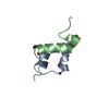
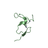
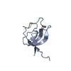
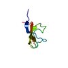
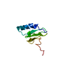
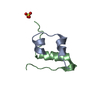
Entry Database : PDB / ID : 4uneTitle Human insulin B26Phe mutant crystal structure INSULIN A CHAIN INSULIN B CHAIN Keywords / Function / homology Function Domain/homology Component
/ / / / / / / / / / / / / / / / / / / / / / / / / / / / / / / / / / / / / / / / / / / / / / / / / / / / / / / / / / / / / / / / / / / / / / / / / / / / / / / / / / / / / / / / / / / / / / / / / / / Biological species HOMO SAPIENS (human)Method / / / Resolution : 1.59 Å Authors Zakova, L. / Klevtikova, E. / Lepsik, M. / Collinsova, M. / Watson, C.J. / Turkenburg, J.P. / Jiracek, J. / Brzozowski, A.M. Journal : Acta Crystallogr.,Sect.D / Year : 2014Title : Human Insulin Analogues Modified at the B26 Site Reveal a Hormone Conformation that is Undetected in the Receptor ComplexAuthors : Zakova, L. / Klevtikova, E. / Lepsik, M. / Collinsova, M. / Watson, C.J. / Turkenburg, J.P. / Jiracek, J. / Brzozowski, A.M. History Deposition May 28, 2014 Deposition site / Processing site Revision 1.0 Oct 15, 2014 Provider / Type Revision 1.1 May 16, 2018 Group / Category / Item Revision 1.2 Jan 10, 2024 Group Data collection / Database references ... Data collection / Database references / Derived calculations / Other / Refinement description Category chem_comp_atom / chem_comp_bond ... chem_comp_atom / chem_comp_bond / database_2 / diffrn_source / pdbx_database_status / pdbx_initial_refinement_model / struct_site Item _database_2.pdbx_DOI / _database_2.pdbx_database_accession ... _database_2.pdbx_DOI / _database_2.pdbx_database_accession / _diffrn_source.pdbx_synchrotron_beamline / _pdbx_database_status.status_code_sf / _struct_site.pdbx_auth_asym_id / _struct_site.pdbx_auth_comp_id / _struct_site.pdbx_auth_seq_id Revision 1.3 Nov 20, 2024 Group / Category / pdbx_modification_feature
Show all Show less
 Movie
Movie Controller


 Open data
 Basic information
 Components
 Keywords
 Function and homology information
 HOMO SAPIENS (human)
 X-RAY DIFFRACTION /
  Authors
 Citation
 Structure visualization
 Downloads & links
 Other downloads
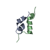


 PDBj
PDBj


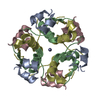
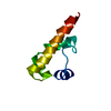
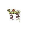
 Assembly


 Mass: 96.063 Da / Num. of mol.: 2 / Source method: obtained synthetically / Formula: SO4
 Mass: 18.015 Da / Num. of mol.: 161 / Source method: isolated from a natural source / Formula: H2O
 Sample preparation
 / Beamline: ID23 / Wavelength: 0.8726
  Processing