 Movie
Movie Controller

[English] 日本語
 Yorodumi
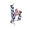
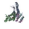
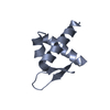
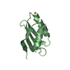
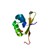
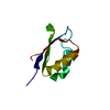
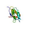
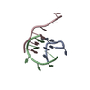
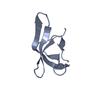
- PDB-1g2y: HNF-1ALPHA DIMERIZATION DOMAIN, WITH SELENOMETHIONINE SUBSTITUED ... -
+ Open data

- Basic information
| Entry | Database: PDB / ID: 1g2y | ||||||
|---|---|---|---|---|---|---|---|
| Title | HNF-1ALPHA DIMERIZATION DOMAIN, WITH SELENOMETHIONINE SUBSTITUED AT LEU 12 | ||||||
 Components Components | HEPATOCYTE NUCLEAR FACTOR 1-ALPHA | ||||||
 Keywords Keywords | TRANSCRIPTION / dimerization domain / four-helix bundle / transcription factor / selenomethionine | ||||||
| Function / homology |  Function and homology information Function and homology informationparaxial mesoderm formation / apoptotic nuclear changes / regulation of NADP metabolic process / renal D-glucose absorption / cellular response to rapamycin / regulation of hormone secretion / reproductive structure development / bile acid biosynthetic process / reverse cholesterol transport / cellular response to L-leucine ...paraxial mesoderm formation / apoptotic nuclear changes / regulation of NADP metabolic process / renal D-glucose absorption / cellular response to rapamycin / regulation of hormone secretion / reproductive structure development / bile acid biosynthetic process / reverse cholesterol transport / cellular response to L-leucine / pancreas development / pronucleus / negative regulation of miRNA processing / regulation of Wnt signaling pathway / embryonic limb morphogenesis / heme biosynthetic process / insulin secretion / positive regulation of mitochondrial membrane potential / : / bile acid and bile salt transport / positive regulation of ATP biosynthetic process / bone resorption / photoreceptor outer segment / blastocyst development / positive regulation of transcription initiation by RNA polymerase II / fatty acid transport / response to glucose / cholesterol metabolic process / placenta development / transcription coregulator binding / liver development / cellular response to glucose stimulus / transcription coactivator binding / fatty acid biosynthetic process / positive regulation of insulin secretion / intracellular protein localization / glucose homeostasis / response to oxidative stress / double-stranded DNA binding / DNA-binding transcription activator activity, RNA polymerase II-specific / transcription regulator complex / transcription by RNA polymerase II / sequence-specific DNA binding / DNA-binding transcription factor activity, RNA polymerase II-specific / positive regulation of phosphatidylinositol 3-kinase/protein kinase B signal transduction / protein dimerization activity / transcription cis-regulatory region binding / RNA polymerase II cis-regulatory region sequence-specific DNA binding / chromatin remodeling / DNA-binding transcription factor activity / protein heterodimerization activity / chromatin binding / positive regulation of gene expression / regulation of transcription by RNA polymerase II / regulation of DNA-templated transcription / negative regulation of apoptotic process / positive regulation of DNA-templated transcription / chromatin / negative regulation of transcription by RNA polymerase II / protein homodimerization activity / positive regulation of transcription by RNA polymerase II / protein-containing complex / DNA binding / identical protein binding / nucleus / cytoplasm Similarity search - Function | ||||||
| Method |  X-RAY DIFFRACTION / SYNCHROTRON / MAD / Resolution: 1 Å X-RAY DIFFRACTION / SYNCHROTRON / MAD / Resolution: 1 Å | ||||||
 Authors Authors | Rose, R.B. / Endrizzi, J.A. / Cronk, J.D. / Holton, J. / Alber, T. | ||||||
 Citation Citation | Journal: Biochemistry / Year: 2000 Title: High-resolution structure of the HNF-1alpha dimerization domain. Authors: Rose, R.B. / Endrizzi, J.A. / Cronk, J.D. / Holton, J. / Alber, T. #1: Journal: Nat.Struct.Biol. / Year: 2000Title: Structural basis of dimerization, coactivator recognition and MODY3 mutations in HNF-1alpha Authors: Rose, R.B. / Bayle, J.H. / Endrizzi, J.A. / Cronk, J.D. / Crabtree, G.R. / Alber, T. | ||||||
| History |
|
- Structure visualization
| Structure viewer | Molecule: MolmilJmol/JSmol |
|---|
- Downloads & links
-Download
| PDBx/mmCIF format | 1g2y.cif.gz | 36.1 KB | Display | PDBx/mmCIF format |
|---|---|---|---|---|
| PDB format | pdb1g2y.ent.gz | 26.5 KB | Display | PDB format |
| PDBx/mmJSON format | 1g2y.json.gz | Tree view | PDBx/mmJSON format | |
| Others |  Other downloads Other downloads |
-Validation report
| Arichive directory | https://data.pdbj.org/pub/pdb/validation_reports/g2/1g2yftp://data.pdbj.org/pub/pdb/validation_reports/g2/1g2y | HTTPS FTP |
|---|
-Related structure data


-Links
 PDBj
PDBj


- Assembly
| Deposited unit | 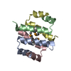
| ||||||||
|---|---|---|---|---|---|---|---|---|---|
| 1 | 
| ||||||||
| 2 | 
| ||||||||
| 3 |
| ||||||||
| 4 | 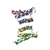
| ||||||||
| Unit cell |
| ||||||||
| Details | There are two HNF-1alpha dimers in the asymmetric unit: monomers A and C, and monomers B and D. |
-Components
| #1: Protein/peptide | Mass: 3451.884 Da / Num. of mol.: 4 / Fragment: DIMERIZATION DOMAIN, RESIDUES 1-32 / Mutation: L12(MSE) / Source method: obtained synthetically Details: This peptide was chemically synthesized. The sequence of this peptide naturally occurs in mouse (Mus musculus), with a point mutation at position 12. References: UniProt: P22361 #2: Water | ChemComp-HOH / |  Mass: 18.015 Da / Num. of mol.: 176 / Source method: isolated from a natural source / Formula: H2O Mass: 18.015 Da / Num. of mol.: 176 / Source method: isolated from a natural source / Formula: H2OHas protein modification | Y | |
|---|
-Experimental details
-Experiment
| Experiment | Method: X-RAY DIFFRACTION / Number of used crystals: 1 |
|---|
- Sample preparation
| Crystal | Density Matthews: 2.19 Å3/Da / Density % sol: 43.83 % | ||||||||||||||||||||||||||||||
|---|---|---|---|---|---|---|---|---|---|---|---|---|---|---|---|---|---|---|---|---|---|---|---|---|---|---|---|---|---|---|---|
| Crystal grow | Temperature: 277 K / Method: vapor diffusion, hanging drop / pH: 8.5 Details: PEG 4000, Tris-HCl, lithium sulphate, pH 8.5, VAPOR DIFFUSION, HANGING DROP, temperature 277.0K | ||||||||||||||||||||||||||||||
| Crystal grow | *PLUS Method: vapor diffusion | ||||||||||||||||||||||||||||||
| Components of the solutions | *PLUS
|
-Data collection
| Diffraction | Mean temperature: 200 K | |||||||||||||||
|---|---|---|---|---|---|---|---|---|---|---|---|---|---|---|---|---|
| Diffraction source | Source: SYNCHROTRON / Site: ALS  / Beamline: 5.0.2 / Wavelength: 0.95372, 0.97957, 0.9798, 1.07812 / Beamline: 5.0.2 / Wavelength: 0.95372, 0.97957, 0.9798, 1.07812 | |||||||||||||||
| Detector | Type: ADSC QUANTUM 4 / Detector: CCD / Date: Oct 28, 1999 / Details: Double crystal | |||||||||||||||
| Radiation | Monochromator: double crystal / Protocol: MAD / Monochromatic (M) / Laue (L): M / Scattering type: x-ray | |||||||||||||||
| Radiation wavelength |
| |||||||||||||||
| Reflection | Resolution: 1→30.9 Å / Num. all: 55221 / Num. obs: 55221 / % possible obs: 86.4 % / Observed criterion σ(F): 2 / Observed criterion σ(I): 2 / Redundancy: 2.8 % / Biso Wilson estimate: 7.4 Å2 / Rmerge(I) obs: 0.049 / Net I/σ(I): 16.8 | |||||||||||||||
| Reflection shell | Resolution: 1→1.05 Å / Redundancy: 2.8 % / Rmerge(I) obs: 0.101 / Mean I/σ(I) obs: 9.2 / Num. unique all: 55221 / % possible all: 73.1 | |||||||||||||||
| Reflection | *PLUS | |||||||||||||||
| Reflection shell | *PLUS % possible obs: 73.1 % |
- Processing
| Software |
| ||||||||||||||||||||||||||||||||||||||||
|---|---|---|---|---|---|---|---|---|---|---|---|---|---|---|---|---|---|---|---|---|---|---|---|---|---|---|---|---|---|---|---|---|---|---|---|---|---|---|---|---|---|
| Refinement | Method to determine structure: MAD Starting model: wARP model Resolution: 1→30.88 Å / Rfactor Rfree error: 0.005 / Data cutoff high absF: 499172.87 / Data cutoff low absF: 0 / Isotropic thermal model: RESTRAINED / Cross valid method: THROUGHOUT / σ(F): 0 / σ(I): 0 / Stereochemistry target values: Engh & Huber
| ||||||||||||||||||||||||||||||||||||||||
| Solvent computation | Solvent model: FLAT MODEL / Bsol: 98.59 Å2 / ksol: 0.372 e/Å3 | ||||||||||||||||||||||||||||||||||||||||
| Displacement parameters | Biso mean: 16 Å2
| ||||||||||||||||||||||||||||||||||||||||
| Refine analyze |
| ||||||||||||||||||||||||||||||||||||||||
| Refinement step | Cycle: LAST / Resolution: 1→30.88 Å
| ||||||||||||||||||||||||||||||||||||||||
| Refine LS restraints |
| ||||||||||||||||||||||||||||||||||||||||
| LS refinement shell | Resolution: 1→1.06 Å / Rfactor Rfree error: 0.025 / Total num. of bins used: 6
| ||||||||||||||||||||||||||||||||||||||||
| Xplor file |
| ||||||||||||||||||||||||||||||||||||||||
| Software | *PLUS Name: CNS / Classification: refinement | ||||||||||||||||||||||||||||||||||||||||
| Refinement | *PLUS σ(F): 0 / Num. reflection Rfree: 5640 / % reflection Rfree: 9 % | ||||||||||||||||||||||||||||||||||||||||
| Solvent computation | *PLUS | ||||||||||||||||||||||||||||||||||||||||
| Displacement parameters | *PLUS Biso mean: 16 Å2 | ||||||||||||||||||||||||||||||||||||||||
| Refine LS restraints | *PLUS
| ||||||||||||||||||||||||||||||||||||||||
| LS refinement shell | *PLUS Rfactor Rfree: 0.375 / % reflection Rfree: 2.9 % / Rfactor Rwork: 0.398 |
