 Movie
Movie Controller

[English] 日本語
 Yorodumi
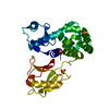
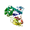
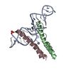
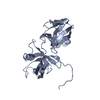
- PDB-1f93: CRYSTAL STRUCTURE OF A COMPLEX BETWEEN THE DIMERIZATION DOMAIN OF... -
+ Open data

- Basic information
| Entry | Database: PDB / ID: 1f93 | ||||||
|---|---|---|---|---|---|---|---|
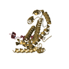
| Title | CRYSTAL STRUCTURE OF A COMPLEX BETWEEN THE DIMERIZATION DOMAIN OF HNF-1 ALPHA AND THE COACTIVATOR DCOH | ||||||
 Components Components |
| ||||||
 Keywords Keywords | TRANSCRIPTION / four-helix bundle / transcriptional activator-coactivator complex / dimerization domain | ||||||
| Function / homology |  Function and homology information Function and homology informationregulation of protein binding / Phenylalanine metabolism / 4a-hydroxytetrahydrobiopterin dehydratase / 4-alpha-hydroxytetrahydrobiopterin dehydratase activity / paraxial mesoderm formation / : / apoptotic nuclear changes / regulation of NADP metabolic process / renal D-glucose absorption / phenylalanine 4-monooxygenase activity ...regulation of protein binding / Phenylalanine metabolism / 4a-hydroxytetrahydrobiopterin dehydratase / 4-alpha-hydroxytetrahydrobiopterin dehydratase activity / paraxial mesoderm formation / : / apoptotic nuclear changes / regulation of NADP metabolic process / renal D-glucose absorption / phenylalanine 4-monooxygenase activity / cellular response to rapamycin / regulation of hormone secretion / reproductive structure development / bile acid biosynthetic process / tetrahydrobiopterin biosynthetic process / reverse cholesterol transport / cellular response to L-leucine / pancreas development / pronucleus / negative regulation of miRNA processing / regulation of Wnt signaling pathway / embryonic limb morphogenesis / heme biosynthetic process / insulin secretion / positive regulation of mitochondrial membrane potential / : / bile acid and bile salt transport / positive regulation of ATP biosynthetic process / photoreceptor outer segment / bone resorption / blastocyst development / positive regulation of transcription initiation by RNA polymerase II / fatty acid transport / response to glucose / cholesterol metabolic process / placenta development / transcription coregulator binding / liver development / cellular response to glucose stimulus / transcription coactivator binding / fatty acid biosynthetic process / positive regulation of insulin secretion / intracellular protein localization / glucose homeostasis / response to oxidative stress / double-stranded DNA binding / DNA-binding transcription activator activity, RNA polymerase II-specific / transcription regulator complex / transcription by RNA polymerase II / sequence-specific DNA binding / DNA-binding transcription factor activity, RNA polymerase II-specific / transcription coactivator activity / positive regulation of phosphatidylinositol 3-kinase/protein kinase B signal transduction / protein dimerization activity / transcription cis-regulatory region binding / RNA polymerase II cis-regulatory region sequence-specific DNA binding / chromatin remodeling / DNA-binding transcription factor activity / protein heterodimerization activity / chromatin binding / positive regulation of gene expression / regulation of transcription by RNA polymerase II / regulation of DNA-templated transcription / negative regulation of apoptotic process / positive regulation of DNA-templated transcription / chromatin / negative regulation of transcription by RNA polymerase II / protein homodimerization activity / positive regulation of transcription by RNA polymerase II / protein-containing complex / DNA binding / nucleoplasm / identical protein binding / nucleus / cytoplasm / cytosol Similarity search - Function | ||||||
| Biological species |  | ||||||
| Method |  X-RAY DIFFRACTION / SYNCHROTRON / Resolution: 2.6 Å X-RAY DIFFRACTION / SYNCHROTRON / Resolution: 2.6 Å | ||||||
 Authors Authors | Rose, R.B. / Bayle, J.H. / Endrizzi, J.A. / Cronk, J.D. / Crabtree, G.R. / Alber, T. | ||||||
 Citation Citation | Journal: Nat.Struct.Biol. / Year: 2000 Title: Structural basis of dimerization, coactivator recognition and MODY3 mutations in HNF-1alpha. Authors: Rose, R.B. / Bayle, J.H. / Endrizzi, J.A. / Cronk, J.D. / Crabtree, G.R. / Alber, T. #1: Journal: Science / Year: 1995Title: Crystal structure of DCoH, a bifunctional, protein-binding transcriptional coactivator Authors: Endrizzi, J.A. / Cronk, J.D. / Weidong, W. / Crabtree, G.R. / Alber, T. #2: Journal: Protein Sci. / Year: 1996Title: High-resolution structures of the bifunctional enzyme and transcriptional coactivator DCoH and its complex with a product analogue Authors: Cronk, J.D. / Endrizzi, J.A. / Alber, T. | ||||||
| History |
|
- Structure visualization
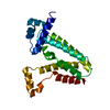
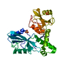
| Structure viewer | Molecule: MolmilJmol/JSmol |
|---|
- Downloads & links
-Download
| PDBx/mmCIF format | 1f93.cif.gz | 112.5 KB | Display | PDBx/mmCIF format |
|---|---|---|---|---|
| PDB format | pdb1f93.ent.gz | 88.5 KB | Display | PDB format |
| PDBx/mmJSON format | 1f93.json.gz | Tree view | PDBx/mmJSON format | |
| Others |  Other downloads Other downloads |
-Validation report
| Arichive directory | https://data.pdbj.org/pub/pdb/validation_reports/f9/1f93ftp://data.pdbj.org/pub/pdb/validation_reports/f9/1f93 | HTTPS FTP |
|---|
-Related structure data
| Similar structure data |
|---|

-Links
 PDBj
PDBj


- Assembly
| Deposited unit | 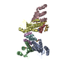
| ||||||||
|---|---|---|---|---|---|---|---|---|---|
| 1 | 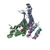
| ||||||||
| 2 | 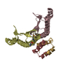
| ||||||||
| Unit cell |
| ||||||||
| Details | The biological assembly is a heterotetramer consisting of a DCoH dimer and an HNF-1 alpha dimerization domain dimer. There are two heterotetramers in the asymmetric unit. |
-Components
| #1: Protein | Mass: 12158.288 Da / Num. of mol.: 4 Source method: isolated from a genetically manipulated source Source: (gene. exp.)  #2: Protein/peptide | Mass: 3386.951 Da / Num. of mol.: 4 / Fragment: DIMERIZATION DOMAIN (RESIDUES 1-32) / Source method: obtained synthetically Details: THIS PEPTIDE WAS CHEMICALLY SYNTHESIZED. THE SEQUENCE OF THIS PEPTIDE NATURALLY OCCURS IN MOUSE (MUS MUSCULUS) References: UniProt: P22361 #3: Water | ChemComp-HOH / |  Mass: 18.015 Da / Num. of mol.: 62 / Source method: isolated from a natural source / Formula: H2O Mass: 18.015 Da / Num. of mol.: 62 / Source method: isolated from a natural source / Formula: H2OHas protein modification | Y | |
|---|
-Experimental details
-Experiment
| Experiment | Method: X-RAY DIFFRACTION / Number of used crystals: 1 |
|---|
- Sample preparation
| Crystal | Density Matthews: 2.3 Å3/Da / Density % sol: 46.61 % | |||||||||||||||||||||||||||||||||||
|---|---|---|---|---|---|---|---|---|---|---|---|---|---|---|---|---|---|---|---|---|---|---|---|---|---|---|---|---|---|---|---|---|---|---|---|---|
| Crystal grow | Temperature: 277 K / Method: vapor diffusion, hanging drop / pH: 5 Details: PEG 8000, potassium succinate, pH 5.0, VAPOR DIFFUSION, HANGING DROP, temperature 277.0K | |||||||||||||||||||||||||||||||||||
| Crystal grow | *PLUS Temperature: 4 ℃ / Method: vapor diffusion / Details: used microseeding | |||||||||||||||||||||||||||||||||||
| Components of the solutions | *PLUS
|
-Data collection
| Diffraction | Mean temperature: 100 K |
|---|---|
| Diffraction source | Source: SYNCHROTRON / Site: SSRL  / Beamline: BL1-5 / Wavelength: 1.0688 / Beamline: BL1-5 / Wavelength: 1.0688 |
| Detector | Type: ADSC QUANTUM / Detector: CCD / Date: Jan 1, 1998 |
| Radiation | Protocol: SINGLE WAVELENGTH / Monochromatic (M) / Laue (L): M / Scattering type: x-ray |
| Radiation wavelength | Wavelength: 1.0688 Å / Relative weight: 1 |
| Reflection | Resolution: 2.6→20 Å / Num. all: 59400 / Num. obs: 16500 / % possible obs: 95 % / Observed criterion σ(F): 4 / Observed criterion σ(I): 2 / Redundancy: 3.6 % / Biso Wilson estimate: 43.3 Å2 / Rmerge(I) obs: 0.06 / Net I/σ(I): 22 |
| Reflection shell | Resolution: 2.6→2.67 Å / Redundancy: 3.4 % / Rmerge(I) obs: 0.3 / Num. unique all: 1800 / % possible all: 95 |
| Reflection | *PLUS % possible obs: 95 % / Num. measured all: 59400 / Rmerge(I) obs: 0.057 |
| Reflection shell | *PLUS % possible obs: 95 % / Rmerge(I) obs: 0.243 / Mean I/σ(I) obs: 2.6 |
- Processing
| Software |
| ||||||||||||||||||||||||||||||||||||||||||||
|---|---|---|---|---|---|---|---|---|---|---|---|---|---|---|---|---|---|---|---|---|---|---|---|---|---|---|---|---|---|---|---|---|---|---|---|---|---|---|---|---|---|---|---|---|---|
| Refinement | Resolution: 2.6→20.1 Å / Rfactor Rfree error: 0.008 / Data cutoff high absF: 912032.47 / Data cutoff low absF: 0 / Isotropic thermal model: RESTRAINED / Cross valid method: THROUGHOUT / σ(F): 0 / σ(I): 0 / Stereochemistry target values: Engh and Huber / Details: used non-crystallographic symmetry
| ||||||||||||||||||||||||||||||||||||||||||||
| Solvent computation | Solvent model: FLAT MODEL / Bsol: 55.35 Å2 / ksol: 0.36 e/Å3 | ||||||||||||||||||||||||||||||||||||||||||||
| Displacement parameters | Biso mean: 58.7 Å2
| ||||||||||||||||||||||||||||||||||||||||||||
| Refine analyze |
| ||||||||||||||||||||||||||||||||||||||||||||
| Refinement step | Cycle: LAST / Resolution: 2.6→20.1 Å
| ||||||||||||||||||||||||||||||||||||||||||||
| Refine LS restraints |
| ||||||||||||||||||||||||||||||||||||||||||||
| LS refinement shell | Resolution: 2.6→2.76 Å / Rfactor Rfree error: 0.024 / Total num. of bins used: 6
| ||||||||||||||||||||||||||||||||||||||||||||
| Xplor file |
| ||||||||||||||||||||||||||||||||||||||||||||
| Software | *PLUS Name: CNS / Version: 0.4 / Classification: refinement | ||||||||||||||||||||||||||||||||||||||||||||
| Refinement | *PLUS σ(F): 0 / % reflection Rfree: 7.8 % | ||||||||||||||||||||||||||||||||||||||||||||
| Solvent computation | *PLUS | ||||||||||||||||||||||||||||||||||||||||||||
| Displacement parameters | *PLUS Biso mean: 58.7 Å2 | ||||||||||||||||||||||||||||||||||||||||||||
| Refine LS restraints | *PLUS
| ||||||||||||||||||||||||||||||||||||||||||||
| LS refinement shell | *PLUS Rfactor Rfree: 0.371 / % reflection Rfree: 8 % / Rfactor Rwork: 0.31 |
