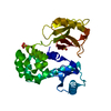
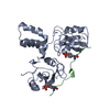
TRANSPORT PROTEIN / MCU / mitochondrial calcium uniporter / Calcium channel
Function / homology
Function and homology information
uniporter activity / uniplex complex / positive regulation of mitochondrial calcium ion concentration / Processing of SMDT1 / Mitochondrial calcium ion transport / mitochondrial calcium ion transmembrane transport / mitochondrial calcium ion homeostasis / calcium import into the mitochondrion / cellular response to calcium ion starvation / positive regulation of neutrophil chemotaxis ...uniporter activity / uniplex complex / positive regulation of mitochondrial calcium ion concentration / Processing of SMDT1 / Mitochondrial calcium ion transport / mitochondrial calcium ion transmembrane transport / mitochondrial calcium ion homeostasis / calcium import into the mitochondrion / cellular response to calcium ion starvation / positive regulation of neutrophil chemotaxis / positive regulation of mitochondrial fission / protein complex oligomerization / viral release from host cell by cytolysis / peptidoglycan catabolic process / calcium channel complex / generation of precursor metabolites and energy / calcium-mediated signaling / cell wall macromolecule catabolic process / calcium channel activity / positive regulation of insulin secretion / lysozyme / lysozyme activity / cell migration / glucose homeostasis / host cell cytoplasm / defense response to bacterium / cell population proliferation / mitochondrial inner membrane / mitochondrion / identical protein binding Similarity search - Function
In the structure databanks used in Yorodumi, some data are registered as the other names, "COVID-19 virus" and "2019-nCoV". Here are the details of the virus and the list of structure data.
Jan 31, 2019. EMDB accession codes are about to change! (news from PDBe EMDB page)
EMDB accession codes are about to change! (news from PDBe EMDB page)
The allocation of 4 digits for EMDB accession codes will soon come to an end. Whilst these codes will remain in use, new EMDB accession codes will include an additional digit and will expand incrementally as the available range of codes is exhausted. The current 4-digit format prefixed with “EMD-” (i.e. EMD-XXXX) will advance to a 5-digit format (i.e. EMD-XXXXX), and so on. It is currently estimated that the 4-digit codes will be depleted around Spring 2019, at which point the 5-digit format will come into force.
The EM Navigator/Yorodumi systems omit the EMD- prefix.
Related info.:Q: What is EMD? / ID/Accession-code notation in Yorodumi/EM Navigator
Yorodumi is a browser for structure data from EMDB, PDB, SASBDB, etc.
This page is also the successor to EM Navigator detail page, and also detail information page/front-end page for Omokage search.
The word "yorodu" (or yorozu) is an old Japanese word meaning "ten thousand". "mi" (miru) is to see.
Related info.:EMDB / PDB / SASBDB / Comparison of 3 databanks / Yorodumi Search / Aug 31, 2016. New EM Navigator & Yorodumi / Yorodumi Papers / Jmol/JSmol / Function and homology information / Changes in new EM Navigator and Yorodumi
 Movie
Movie Controller

 Yorodumi
 Open data
 Basic information
 Components
 Keywords
 Function and homology information
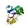
 Enterobacteria phage T4 (virus)
 Homo sapiens (human)
 X-RAY DIFFRACTION /
  Authors
 Citation
 Structure visualization
 Downloads & links
 Other downloads
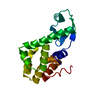


 PDBj
PDBj


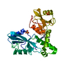
 Assembly


 Mass: 96.063 Da / Num. of mol.: 4 / Source method: obtained synthetically / Formula: SO4
 Sample preparation
 / Beamline: 5C (4A) / Wavelength: 0.9795 Å
 Processing