 Movie
Movie Controller

[English] 日本語
 Yorodumi
- PDB-5bz6: Crystal structure of the N-terminal domain single mutant (S92A) o... -
+ Open data

- Basic information
| Entry | Database: PDB / ID: 5bz6 | ||||||
|---|---|---|---|---|---|---|---|
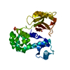
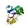
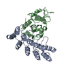
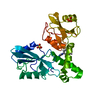
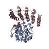
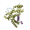
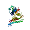
| Title | Crystal structure of the N-terminal domain single mutant (S92A) of the human mitochondrial calcium uniporter fused with T4 lysozyme | ||||||
 Components Components | Lysozyme,Calcium uniporter protein, mitochondrial | ||||||
 Keywords Keywords | TRANSPORT PROTEIN / Membrane protein / Calcium channel / Mitochondria | ||||||
| Function / homology |  Function and homology information Function and homology informationuniporter activity / uniplex complex / Processing of SMDT1 / positive regulation of mitochondrial calcium ion concentration / Mitochondrial calcium ion transport / mitochondrial calcium ion transmembrane transport / mitochondrial calcium ion homeostasis / calcium import into the mitochondrion / cellular response to calcium ion starvation / positive regulation of neutrophil chemotaxis ...uniporter activity / uniplex complex / Processing of SMDT1 / positive regulation of mitochondrial calcium ion concentration / Mitochondrial calcium ion transport / mitochondrial calcium ion transmembrane transport / mitochondrial calcium ion homeostasis / calcium import into the mitochondrion / cellular response to calcium ion starvation / positive regulation of neutrophil chemotaxis / positive regulation of mitochondrial fission / protein complex oligomerization / viral release from host cell by cytolysis / peptidoglycan catabolic process / calcium channel complex / generation of precursor metabolites and energy / calcium-mediated signaling / calcium channel activity / cell wall macromolecule catabolic process / positive regulation of insulin secretion / lysozyme / lysozyme activity / cell migration / glucose homeostasis / host cell cytoplasm / cell population proliferation / defense response to bacterium / mitochondrial inner membrane / mitochondrion / identical protein binding Similarity search - Function | ||||||
| Biological species |  Enterobacteria phage T4 (virus) Enterobacteria phage T4 (virus) Homo sapiens (human) Homo sapiens (human) | ||||||
| Method |  X-RAY DIFFRACTION / SYNCHROTRON / MOLECULAR REPLACEMENT / Resolution: 2.75 Å X-RAY DIFFRACTION / SYNCHROTRON / MOLECULAR REPLACEMENT / Resolution: 2.75 Å | ||||||
 Authors Authors | Lee, Y. / Min, C.K. / Kim, T.G. / Song, H.K. / Lim, Y. / Kim, D. / Shin, K. / Kang, M. / Kang, J.Y. / Youn, H.-S. ...Lee, Y. / Min, C.K. / Kim, T.G. / Song, H.K. / Lim, Y. / Kim, D. / Shin, K. / Kang, M. / Kang, J.Y. / Youn, H.-S. / Lee, J.-G. / An, J.Y. / Park, K.R. / Lim, J.J. / Kim, J.H. / Kim, J.H. / Park, Z.Y. / Kim, Y.-S. / Wang, J. / Kim, D.H. / Eom, S.H. | ||||||
 Citation Citation | Journal: Embo Rep. / Year: 2015 Title: Structure and function of the N-terminal domain of the human mitochondrial calcium uniporter. Authors: Lee, Y. / Min, C.K. / Kim, T.G. / Song, H.K. / Lim, Y. / Kim, D. / Shin, K. / Kang, M. / Kang, J.Y. / Youn, H.S. / Lee, J.G. / An, J.Y. / Park, K.R. / Lim, J.J. / Kim, J.H. / Kim, J.H. / ...Authors: Lee, Y. / Min, C.K. / Kim, T.G. / Song, H.K. / Lim, Y. / Kim, D. / Shin, K. / Kang, M. / Kang, J.Y. / Youn, H.S. / Lee, J.G. / An, J.Y. / Park, K.R. / Lim, J.J. / Kim, J.H. / Kim, J.H. / Park, Z.Y. / Kim, Y.S. / Wang, J. / Kim, D.H. / Eom, S.H. | ||||||
| History |
|
- Structure visualization
| Structure viewer | Molecule: MolmilJmol/JSmol |
|---|
- Downloads & links
-Download
| PDBx/mmCIF format | 5bz6.cif.gz | 66 KB | Display | PDBx/mmCIF format |
|---|---|---|---|---|
| PDB format | pdb5bz6.ent.gz | 47.7 KB | Display | PDB format |
| PDBx/mmJSON format | 5bz6.json.gz | Tree view | PDBx/mmJSON format | |
| Others |  Other downloads Other downloads |
-Validation report
| Arichive directory | https://data.pdbj.org/pub/pdb/validation_reports/bz/5bz6ftp://data.pdbj.org/pub/pdb/validation_reports/bz/5bz6 | HTTPS FTP |
|---|
-Related structure data
| Related structure data |  4xsjSC  4xtbC 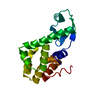 2lzmS S: Starting model for refinement C: citing same article ( |
|---|---|
| Similar structure data |


-Links
 PDBj
PDBj


- Assembly
| Deposited unit | 
| ||||||||
|---|---|---|---|---|---|---|---|---|---|
| 1 |
| ||||||||
| Unit cell |
|
-Components
| #1: Protein | Mass: 29772.135 Da / Num. of mol.: 1 / Mutation: D20N, C54T, C97A, S1092A Source method: isolated from a genetically manipulated source Details: The fusion protein of bacteriophage T4 lysozyme protein (UNP RESIDUES 1-163), LINKER GS, anmitochondrial calcium uniporter N-terminal domain (UNP RESIDUES 75-165) and tags LEHHHHHH Source: (gene. exp.) Enterobacteria phage T4 (virus), (gene. exp.) Homo sapiens (human)Gene: e, T4Tp126, MCU, C10orf42, CCDC109A / Production host:  | ||
|---|---|---|---|
| #2: Chemical | ChemComp-SO4 /   Mass: 96.063 Da / Num. of mol.: 6 / Source method: obtained synthetically / Formula: SO4 Mass: 96.063 Da / Num. of mol.: 6 / Source method: obtained synthetically / Formula: SO4#3: Water | ChemComp-HOH / |  Mass: 18.015 Da / Num. of mol.: 41 / Source method: isolated from a natural source / Formula: H2O Mass: 18.015 Da / Num. of mol.: 41 / Source method: isolated from a natural source / Formula: H2O |
-Experimental details
-Experiment
| Experiment | Method: X-RAY DIFFRACTION |
|---|
- Sample preparation
| Crystal | Density Matthews: 2.98 Å3/Da / Density % sol: 58.68 % |
|---|---|
| Crystal grow | Temperature: 293 K / Method: vapor diffusion, hanging drop / pH: 6.5 Details: 25% PEG 3350, 0.1M Bis-Tris-HCl (pH 6.5), 0.2M ammonium sulfate |
-Data collection
| Diffraction | Mean temperature: 100 K |
|---|---|
| Diffraction source | Source: SYNCHROTRON / Site: PAL/PLS  / Beamline: 5C (4A) / Wavelength: 0.979 Å / Beamline: 5C (4A) / Wavelength: 0.979 Å |
| Detector | Type: ADSC QUANTUM 315r / Detector: CCD / Date: Sep 14, 2013 |
| Radiation | Protocol: SINGLE WAVELENGTH / Monochromatic (M) / Laue (L): M / Scattering type: x-ray |
| Radiation wavelength | Wavelength: 0.979 Å / Relative weight: 1 |
| Reflection | Resolution: 2.75→50 Å / Num. obs: 11171 / % possible obs: 99.47 % / Redundancy: 4.7 % / Net I/σ(I): 12.7 |
- Processing
| Software |
| ||||||||||||||||||||||||||||||||||||||||||||||||||||||||||||||||||||||||||||||||||||||||||||||||||||||||||||||||||||||||||||||||||||||||||||||||||||||||||||||||||||||||||||||||||||||
|---|---|---|---|---|---|---|---|---|---|---|---|---|---|---|---|---|---|---|---|---|---|---|---|---|---|---|---|---|---|---|---|---|---|---|---|---|---|---|---|---|---|---|---|---|---|---|---|---|---|---|---|---|---|---|---|---|---|---|---|---|---|---|---|---|---|---|---|---|---|---|---|---|---|---|---|---|---|---|---|---|---|---|---|---|---|---|---|---|---|---|---|---|---|---|---|---|---|---|---|---|---|---|---|---|---|---|---|---|---|---|---|---|---|---|---|---|---|---|---|---|---|---|---|---|---|---|---|---|---|---|---|---|---|---|---|---|---|---|---|---|---|---|---|---|---|---|---|---|---|---|---|---|---|---|---|---|---|---|---|---|---|---|---|---|---|---|---|---|---|---|---|---|---|---|---|---|---|---|---|---|---|---|---|
| Refinement | Method to determine structure: MOLECULAR REPLACEMENT Starting model: 2LZM, 4XSJ Resolution: 2.75→34.89 Å / Cor.coef. Fo:Fc: 0.947 / Cor.coef. Fo:Fc free: 0.867 / SU B: 11.522 / SU ML: 0.232 / Cross valid method: THROUGHOUT / ESU R Free: 0.334 / Stereochemistry target values: MAXIMUM LIKELIHOOD / Details: HYDROGENS HAVE BEEN ADDED IN THE RIDING POSITIONS
| ||||||||||||||||||||||||||||||||||||||||||||||||||||||||||||||||||||||||||||||||||||||||||||||||||||||||||||||||||||||||||||||||||||||||||||||||||||||||||||||||||||||||||||||||||||||
| Solvent computation | Ion probe radii: 0.8 Å / Shrinkage radii: 0.8 Å / VDW probe radii: 1.2 Å / Solvent model: MASK | ||||||||||||||||||||||||||||||||||||||||||||||||||||||||||||||||||||||||||||||||||||||||||||||||||||||||||||||||||||||||||||||||||||||||||||||||||||||||||||||||||||||||||||||||||||||
| Displacement parameters | Biso mean: 34.21 Å2
| ||||||||||||||||||||||||||||||||||||||||||||||||||||||||||||||||||||||||||||||||||||||||||||||||||||||||||||||||||||||||||||||||||||||||||||||||||||||||||||||||||||||||||||||||||||||
| Refinement step | Cycle: LAST / Resolution: 2.75→34.89 Å
| ||||||||||||||||||||||||||||||||||||||||||||||||||||||||||||||||||||||||||||||||||||||||||||||||||||||||||||||||||||||||||||||||||||||||||||||||||||||||||||||||||||||||||||||||||||||
| Refine LS restraints |
|