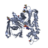
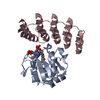
BIOMOLECULE: 1, 2, 3, 4, 5, 6, 7, 8 THIS ENTRY CONTAINS THE CRYSTALLOGRAPHIC ASYMMETRIC UNIT WHICH ...BIOMOLECULE: 1, 2, 3, 4, 5, 6, 7, 8 THIS ENTRY CONTAINS THE CRYSTALLOGRAPHIC ASYMMETRIC UNIT WHICH CONSISTS OF 8 CHAIN(S). AUTHORS STATE THAT THE BIOLOGICAL UNIT OF THIS PROTEIN IS UNKNOWN.
Remark 999
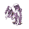
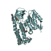
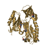
SEQUENCE RESIDUES 1-134 ARE NOT VISIBLE IN THE CRYSTAL STRUCTURE. ACCORDING TO THE AUTHORS, THESE ...SEQUENCE RESIDUES 1-134 ARE NOT VISIBLE IN THE CRYSTAL STRUCTURE. ACCORDING TO THE AUTHORS, THESE RESIDUES WERE APPARENTLY CLEAVED BY SUBTILISIN.
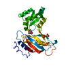
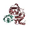
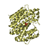
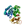
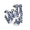
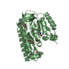
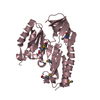
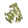
A: Protein TM_1862 B: Protein TM_1862 C: Protein TM_1862 D: Protein TM_1862 E: Protein TM_1862 F: Protein TM_1862 G: Protein TM_1862 H: Protein TM_1862 hetero molecules
Mass: 18.015 Da / Num. of mol.: 1442 / Source method: isolated from a natural source / Formula: H2O
Has protein modification
Y
-
Experimental details
-
Experiment
Experiment
Method: X-RAY DIFFRACTION / Number of used crystals: 1
-
Sample preparation
Crystal
Density Matthews: 2.66 Å3/Da / Density % sol: 53.74 %
Crystal grow
Temperature: 291 K / Method: microbatch under oil / pH: 10 Details: Protein solution: 10 mM Tris-HCl pH 7.5, 100 mM Sodium chloride, 20 micro-g/ml Subtilisin, 5 mM DTT. Reservoir solution: 100 mM CAPS pH 10.0, 40% PEG 4000, 100 mM Sodium thiosulfate, ...Details: Protein solution: 10 mM Tris-HCl pH 7.5, 100 mM Sodium chloride, 20 micro-g/ml Subtilisin, 5 mM DTT. Reservoir solution: 100 mM CAPS pH 10.0, 40% PEG 4000, 100 mM Sodium thiosulfate, MICROBATCH UNDER OIL, temperature 291K
Resolution: 2→2.07 Å / Redundancy: 1.7 % / Rmerge(I) obs: 0.36 / Mean I/σ(I) obs: 3.54 / Rsym value: 0.428 / % possible all: 96
-
Processing
Software
Name
Version
Classification
CNS
1.1
refinement
ADSC
Quantum
datacollection
HKL-2000
datareduction
HKL-2000
datascaling
SnB
phasing
SOLVE
phasing
RESOLVE
phasing
Refinement
Method to determine structure: SAD / Resolution: 2→39.67 Å / Rfactor Rfree error: 0.001 / Data cutoff high absF: 106775.95 / Data cutoff low absF: 0 / Isotropic thermal model: OVERALL / Cross valid method: THROUGHOUT / σ(F): 2 / Stereochemistry target values: Engh & Huber / Details: XtalView program has also been used in refinement
In the structure databanks used in Yorodumi, some data are registered as the other names, "COVID-19 virus" and "2019-nCoV". Here are the details of the virus and the list of structure data.
Jan 31, 2019. EMDB accession codes are about to change! (news from PDBe EMDB page)
EMDB accession codes are about to change! (news from PDBe EMDB page)
The allocation of 4 digits for EMDB accession codes will soon come to an end. Whilst these codes will remain in use, new EMDB accession codes will include an additional digit and will expand incrementally as the available range of codes is exhausted. The current 4-digit format prefixed with “EMD-” (i.e. EMD-XXXX) will advance to a 5-digit format (i.e. EMD-XXXXX), and so on. It is currently estimated that the 4-digit codes will be depleted around Spring 2019, at which point the 5-digit format will come into force.
The EM Navigator/Yorodumi systems omit the EMD- prefix.
Related info.:Q: What is EMD? / ID/Accession-code notation in Yorodumi/EM Navigator
Yorodumi is a browser for structure data from EMDB, PDB, SASBDB, etc.
This page is also the successor to EM Navigator detail page, and also detail information page/front-end page for Omokage search.
The word "yorodu" (or yorozu) is an old Japanese word meaning "ten thousand". "mi" (miru) is to see.
Related info.:EMDB / PDB / SASBDB / Comparison of 3 databanks / Yorodumi Search / Aug 31, 2016. New EM Navigator & Yorodumi / Yorodumi Papers / Jmol/JSmol / Function and homology information / Changes in new EM Navigator and Yorodumi
 Movie
Movie Controller

 Yorodumi
 Open data
 Basic information
 Components
 Keywords
 Function and homology information
 Thermotoga maritima MSB8 (bacteria)
 X-RAY DIFFRACTION /
  Authors
 Citation
 Structure visualization
 Downloads & links
 Other downloads
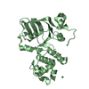


 PDBj
PDBj Assembly
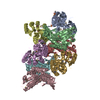


 Mass: 221.317 Da / Num. of mol.: 4 / Source method: obtained synthetically / Formula: C9H19NO3S / Comment: pH buffer*YM
 Mass: 18.015 Da / Num. of mol.: 1442 / Source method: isolated from a natural source / Formula: H2O
 Sample preparation
 / Beamline: X4A / Wavelength: 0.9791 Å
 Processing