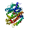
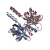
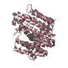
- PDB-1td6: Crystal structure of the conserved hypothetical protein MP506/MPN... -
+
Open data
ID or keywords:
Loading...
-
Basic information
Entry
Database: PDB / ID: 1td6
Title
Crystal structure of the conserved hypothetical protein MP506/MPN330 (gi: 1674200)from Mycoplasma pneumoniae
Components
Hypothetical protein MG237 homolog
Keywords
STRUCTURAL GENOMICS / UNKNOWN FUNCTION / ALPHA HELICAL / PSI / Protein Structure Initiative / Berkeley Structural Genomics Center / BSGC
Function / homology
Function and homology information
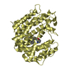
Hypothetical protein mg237 homolog; domain 3 / hypothetical protein mp506/mpn330, domain 1 / hypothetical protein mp506/mpn330, domain 2 / hypothetical protein mp506/mpn330, domain 2 / Uncharacterised protein MG237, central domain / Uncharacterised protein MG237, N-terminal / Protein of unknown function DUF3196 / Protein of unknown function (DUF3196) / hypothetical protein mp506/mpn330, domain 1 / Cyclin A; domain 1 ...Hypothetical protein mg237 homolog; domain 3 / hypothetical protein mp506/mpn330, domain 1 / hypothetical protein mp506/mpn330, domain 2 / hypothetical protein mp506/mpn330, domain 2 / Uncharacterised protein MG237, central domain / Uncharacterised protein MG237, N-terminal / Protein of unknown function DUF3196 / Protein of unknown function (DUF3196) / hypothetical protein mp506/mpn330, domain 1 / Cyclin A; domain 1 / Up-down Bundle / 2-Layer Sandwich / Orthogonal Bundle / Mainly Alpha / Alpha Beta Similarity search - Domain/homology
Mass: 18.015 Da / Num. of mol.: 50 / Source method: isolated from a natural source / Formula: H2O
-
Experimental details
-
Experiment
Experiment
Method: X-RAY DIFFRACTION / Number of used crystals: 1
-
Sample preparation
Crystal
Density Matthews: 2.96 Å3/Da / Density % sol: 58 %
Crystal grow
Temperature: 298 K Method: vapor diffusion, sitting drop (hydra crystallization robot) pH: 8.5 Details: 0.1 M Tris pH 8.5, 1.5 M Lithium Sulfate monohydrate, VAPOR DIFFUSION, SITTING DROP (HYDRA CRYSTALLIZATION ROBOT), temperature 298K
-
Data collection
Diffraction
Mean temperature: 100 K
Diffraction source
Source: SYNCHROTRON / Site: ALS / Beamline: 5.0.2 / Wavelength: 0.9792 Å
In the structure databanks used in Yorodumi, some data are registered as the other names, "COVID-19 virus" and "2019-nCoV". Here are the details of the virus and the list of structure data.
Jan 31, 2019. EMDB accession codes are about to change! (news from PDBe EMDB page)
EMDB accession codes are about to change! (news from PDBe EMDB page)
The allocation of 4 digits for EMDB accession codes will soon come to an end. Whilst these codes will remain in use, new EMDB accession codes will include an additional digit and will expand incrementally as the available range of codes is exhausted. The current 4-digit format prefixed with “EMD-” (i.e. EMD-XXXX) will advance to a 5-digit format (i.e. EMD-XXXXX), and so on. It is currently estimated that the 4-digit codes will be depleted around Spring 2019, at which point the 5-digit format will come into force.
The EM Navigator/Yorodumi systems omit the EMD- prefix.
Related info.:Q: What is EMD? / ID/Accession-code notation in Yorodumi/EM Navigator
Yorodumi is a browser for structure data from EMDB, PDB, SASBDB, etc.
This page is also the successor to EM Navigator detail page, and also detail information page/front-end page for Omokage search.
The word "yorodu" (or yorozu) is an old Japanese word meaning "ten thousand". "mi" (miru) is to see.
Related info.:EMDB / PDB / SASBDB / Comparison of 3 databanks / Yorodumi Search / Aug 31, 2016. New EM Navigator & Yorodumi / Yorodumi Papers / Jmol/JSmol / Function and homology information / Changes in new EM Navigator and Yorodumi
 Movie
Movie Controller

 Yorodumi
 Open data
 Basic information
 Components
 Keywords
 Function and homology information
 Mycoplasma pneumoniae (Filterable agent of primary atypical pneumonia)
 X-RAY DIFFRACTION /
  Authors
 Citation
 Structure visualization
 Downloads & links
 Other downloads
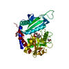


 PDBj
PDBj Assembly
 Mass: 18.015 Da / Num. of mol.: 50 / Source method: isolated from a natural source / Formula: H2O
 Sample preparation
 / Beamline: 5.0.2 / Wavelength: 0.9792 Å
 Processing