 Movie
Movie Controller

[English] 日本語
 Yorodumi
- PDB-4u6r: Crystal structure of human IRE1 cytoplasmic domains in complex wi... -
+ Open data

- Basic information
| Entry | Database: PDB / ID: 4u6r | ||||||
|---|---|---|---|---|---|---|---|
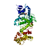
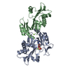
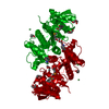
| Title | Crystal structure of human IRE1 cytoplasmic domains in complex with a sulfonamide inhibitor. | ||||||
 Components Components | Serine/threonine-protein kinase/endoribonuclease IRE1 | ||||||
 Keywords Keywords | TRANSFERASE/TRANSFERASE INHIBITOR / Transferase / Kinase / Hydrolase / Endoribonuclease / TRANSFERASE-TRANSFERASE INHIBITOR complex | ||||||
| Function / homology |  Function and homology information Function and homology informationAIP1-IRE1 complex / Ire1 complex / mRNA splicing, via endonucleolytic cleavage and ligation / IRE1alpha activates chaperones / IRE1-TRAF2-ASK1 complex / insulin metabolic process / positive regulation of endoplasmic reticulum unfolded protein response / Hydrolases; Acting on ester bonds; Endoribonucleases producing 5'-phosphomonoesters / platelet-derived growth factor receptor binding / endothelial cell proliferation ...AIP1-IRE1 complex / Ire1 complex / mRNA splicing, via endonucleolytic cleavage and ligation / IRE1alpha activates chaperones / IRE1-TRAF2-ASK1 complex / insulin metabolic process / positive regulation of endoplasmic reticulum unfolded protein response / Hydrolases; Acting on ester bonds; Endoribonucleases producing 5'-phosphomonoesters / platelet-derived growth factor receptor binding / endothelial cell proliferation / nuclear inner membrane / IRE1-RACK1-PP2A complex / IRE1-mediated unfolded protein response / mRNA catabolic process / negative regulation of intrinsic apoptotic signaling pathway / intrinsic apoptotic signaling pathway in response to endoplasmic reticulum stress / cellular response to vascular endothelial growth factor stimulus / cellular response to unfolded protein / regulation of macroautophagy / positive regulation of vascular associated smooth muscle cell proliferation / endoplasmic reticulum unfolded protein response / RNA endonuclease activity / Hsp70 protein binding / response to endoplasmic reticulum stress / positive regulation of RNA splicing / cellular response to glucose stimulus / Hsp90 protein binding / ADP binding / positive regulation of JNK cascade / cellular response to hydrogen peroxide / : / protein phosphorylation / non-specific serine/threonine protein kinase / protein serine kinase activity / hydrolase activity / protein serine/threonine kinase activity / endoplasmic reticulum membrane / magnesium ion binding / enzyme binding / endoplasmic reticulum / protein homodimerization activity / mitochondrion / ATP binding / identical protein binding / cytoplasm Similarity search - Function | ||||||
| Biological species |  Homo sapiens (human) Homo sapiens (human) | ||||||
| Method |  X-RAY DIFFRACTION / MOLECULAR REPLACEMENT / Resolution: 2.5 Å X-RAY DIFFRACTION / MOLECULAR REPLACEMENT / Resolution: 2.5 Å | ||||||
 Authors Authors | Mohr, C. | ||||||
 Citation Citation | Journal: Acs Med.Chem.Lett. / Year: 2015 Title: Unfolded Protein Response in Cancer: IRE1 alpha Inhibition by Selective Kinase Ligands Does Not Impair Tumor Cell Viability. Authors: Harrington, P.E. / Biswas, K. / Malwitz, D. / Tasker, A.S. / Mohr, C. / Andrews, K.L. / Dellamaggiore, K. / Kendall, R. / Beckmann, H. / Jaeckel, P. / Materna-Reichelt, S. / Allen, J.R. / Lipford, J.R. | ||||||
| History |
|
- Structure visualization
| Structure viewer | Molecule: MolmilJmol/JSmol |
|---|
- Downloads & links
-Download
| PDBx/mmCIF format | 4u6r.cif.gz | 184.5 KB | Display | PDBx/mmCIF format |
|---|---|---|---|---|
| PDB format | pdb4u6r.ent.gz | 143 KB | Display | PDB format |
| PDBx/mmJSON format | 4u6r.json.gz | Tree view | PDBx/mmJSON format | |
| Others |  Other downloads Other downloads |
-Validation report
| Arichive directory | https://data.pdbj.org/pub/pdb/validation_reports/u6/4u6rftp://data.pdbj.org/pub/pdb/validation_reports/u6/4u6r | HTTPS FTP |
|---|
-Related structure data
| Related structure data |  4u79C 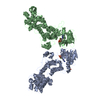 3p23S C: citing same article ( S: Starting model for refinement |
|---|---|
| Similar structure data |


-Links
 PDBj
PDBj- Assembly
| Deposited unit | 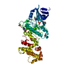
| ||||||||
|---|---|---|---|---|---|---|---|---|---|
| 1 |
| ||||||||
| Unit cell |
|
-Components
-Protein , 1 types, 1 molecules A
| #1: Protein | Mass: 52374.629 Da / Num. of mol.: 1 / Fragment: UNP residues 547-977 Source method: isolated from a genetically manipulated source Source: (gene. exp.) Homo sapiens (human) / Gene: ERN1, IRE1 / Production host:  Trichoplusia ni (cabbage looper) Trichoplusia ni (cabbage looper)References: UniProt: O75460, non-specific serine/threonine protein kinase |
|---|
-Non-polymers , 5 types, 90 molecules 


| #2: Chemical | ChemComp-3E4 /  Mass: 615.145 Da / Num. of mol.: 1 / Source method: obtained synthetically / Formula: C32H31ClN6O3S Mass: 615.145 Da / Num. of mol.: 1 / Source method: obtained synthetically / Formula: C32H31ClN6O3S | ||||||
|---|---|---|---|---|---|---|---|
| #3: Chemical | ChemComp-SO4 /  Mass: 96.063 Da / Num. of mol.: 5 / Source method: obtained synthetically / Formula: SO4 Mass: 96.063 Da / Num. of mol.: 5 / Source method: obtained synthetically / Formula: SO4#4: Chemical | ChemComp-EDO / |  Mass: 62.068 Da / Num. of mol.: 1 / Source method: obtained synthetically / Formula: C2H6O2 Mass: 62.068 Da / Num. of mol.: 1 / Source method: obtained synthetically / Formula: C2H6O2#5: Chemical | ChemComp-EPE / |  Mass: 238.305 Da / Num. of mol.: 1 / Source method: obtained synthetically / Formula: C8H18N2O4S / Comment: pH buffer*YM Mass: 238.305 Da / Num. of mol.: 1 / Source method: obtained synthetically / Formula: C8H18N2O4S / Comment: pH buffer*YM#6: Water | ChemComp-HOH / | Mass: 18.015 Da / Num. of mol.: 82 / Source method: isolated from a natural source / Formula: H2O |
-Details
| Sequence details | SER TO THR CONFLICT AT THIS POSITION IN UNP ENTRY O75460 |
|---|
-Experimental details
-Experiment
| Experiment | Method: X-RAY DIFFRACTION / Number of used crystals: 1 |
|---|
- Sample preparation
| Crystal | Density Matthews: 2.91 Å3/Da / Density % sol: 57.75 % |
|---|---|
| Crystal grow | Temperature: 293 K / Method: vapor diffusion, hanging drop / pH: 7.5 Details: 0.2M Ammonium sulfate, 0.1M HEPES, 25% PEG3350, pH7.5 |
-Data collection
| Diffraction | Mean temperature: 100 K |
|---|---|
| Diffraction source | Source: ROTATING ANODE / Type: RIGAKU FR-E SUPERBRIGHT / Wavelength: 1.5418 Å |
| Detector | Type: RIGAKU SATURN 944 / Detector: CCD / Date: Sep 1, 2011 |
| Radiation | Monochromator: VariMax HF / Protocol: SINGLE WAVELENGTH / Monochromatic (M) / Laue (L): M / Scattering type: x-ray |
| Radiation wavelength | Wavelength: 1.5418 Å / Relative weight: 1 |
| Reflection | Resolution: 2.5→29.47 Å / Num. obs: 21786 / % possible obs: 98.8 % / Redundancy: 7.3 % / Rmerge(I) obs: 0.089 / Net I/σ(I): 9.7 |
| Reflection shell | Resolution: 2.5→2.59 Å / Redundancy: 7.4 % / Rmerge(I) obs: 0.457 / Mean I/σ(I) obs: 4.52 / % possible all: 97.7 |
- Processing
| Software | Name: REFMAC / Version: 5.8.0049 / Classification: refinement | ||||||||||||||||||||||||||||||||||||||||||||||||||||||||||||||||||||||||||||||||||||||||||||||||||||||||||||||||||||||||||||||||||||||||||||||||||||||||||||||||||||||||||||||||||||||
|---|---|---|---|---|---|---|---|---|---|---|---|---|---|---|---|---|---|---|---|---|---|---|---|---|---|---|---|---|---|---|---|---|---|---|---|---|---|---|---|---|---|---|---|---|---|---|---|---|---|---|---|---|---|---|---|---|---|---|---|---|---|---|---|---|---|---|---|---|---|---|---|---|---|---|---|---|---|---|---|---|---|---|---|---|---|---|---|---|---|---|---|---|---|---|---|---|---|---|---|---|---|---|---|---|---|---|---|---|---|---|---|---|---|---|---|---|---|---|---|---|---|---|---|---|---|---|---|---|---|---|---|---|---|---|---|---|---|---|---|---|---|---|---|---|---|---|---|---|---|---|---|---|---|---|---|---|---|---|---|---|---|---|---|---|---|---|---|---|---|---|---|---|---|---|---|---|---|---|---|---|---|---|---|
| Refinement | Method to determine structure: MOLECULAR REPLACEMENT Starting model: 3P23 Resolution: 2.5→29.47 Å / Cor.coef. Fo:Fc: 0.939 / Cor.coef. Fo:Fc free: 0.913 / SU B: 15.495 / SU ML: 0.173 / Cross valid method: THROUGHOUT / ESU R: 0.37 / ESU R Free: 0.258 Stereochemistry target values: MAXIMUM LIKELIHOOD WITH PHASES Details: HYDROGENS HAVE BEEN ADDED IN THE RIDING POSITIONS
| ||||||||||||||||||||||||||||||||||||||||||||||||||||||||||||||||||||||||||||||||||||||||||||||||||||||||||||||||||||||||||||||||||||||||||||||||||||||||||||||||||||||||||||||||||||||
| Solvent computation | Ion probe radii: 0.8 Å / Shrinkage radii: 0.8 Å / VDW probe radii: 1.2 Å / Solvent model: MASK | ||||||||||||||||||||||||||||||||||||||||||||||||||||||||||||||||||||||||||||||||||||||||||||||||||||||||||||||||||||||||||||||||||||||||||||||||||||||||||||||||||||||||||||||||||||||
| Displacement parameters | Biso mean: 43.874 Å2
| ||||||||||||||||||||||||||||||||||||||||||||||||||||||||||||||||||||||||||||||||||||||||||||||||||||||||||||||||||||||||||||||||||||||||||||||||||||||||||||||||||||||||||||||||||||||
| Refinement step | Cycle: 1 / Resolution: 2.5→29.47 Å
| ||||||||||||||||||||||||||||||||||||||||||||||||||||||||||||||||||||||||||||||||||||||||||||||||||||||||||||||||||||||||||||||||||||||||||||||||||||||||||||||||||||||||||||||||||||||
| Refine LS restraints |
|