Mass: 18.015 Da / Num. of mol.: 58 / Source method: isolated from a natural source / Formula: H2O
Has protein modification
Y
-
Experimental details
-
Experiment
Experiment
Method: X-RAY DIFFRACTION / Number of used crystals: 1
-
Sample preparation
Crystal
Density Matthews: 3.2 Å3/Da / Density % sol: 61.6 %
Crystal grow
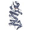
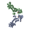
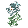
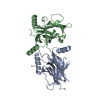
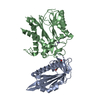
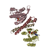
Temperature: 293 K / Method: vapor diffusion, sitting drop / pH: 7.5 Details: The complex of pIRE1a (547-977) with staurosporine was prepared by mixing the protein with 0.5 mM staurosporine and incubated overnight on ice. The crystals were grown at 20C by vapor ...Details: The complex of pIRE1a (547-977) with staurosporine was prepared by mixing the protein with 0.5 mM staurosporine and incubated overnight on ice. The crystals were grown at 20C by vapor diffusion in sitting drop containing 2 ul of protein (13 mg/ml in 50 mM Hepes, pH 7.5, 200 mM NaCl, 5 mM DTT, 1 mM EDTA) and 2ul reservoir solution containing PEG 300 (30%-40%), 100 mM Hepes pH 7.5, 200 mM KCl. Small hexagonal plates appeared over 5-10 days and reached full size (0.05 X 0.75 X 0.15 mm) in ~20 days. The crystals were flash-frozen in liquid N2 directly from the crystallization drop. All diffraction data was collected at the Advanced Photon Source, Argonne National Laboratories, Life Sciences CAT, Sector 21
-
Data collection
Diffraction
Mean temperature: 100 K / Ambient temp details: single crystal
In the structure databanks used in Yorodumi, some data are registered as the other names, "COVID-19 virus" and "2019-nCoV". Here are the details of the virus and the list of structure data.
Jan 31, 2019. EMDB accession codes are about to change! (news from PDBe EMDB page)
EMDB accession codes are about to change! (news from PDBe EMDB page)
The allocation of 4 digits for EMDB accession codes will soon come to an end. Whilst these codes will remain in use, new EMDB accession codes will include an additional digit and will expand incrementally as the available range of codes is exhausted. The current 4-digit format prefixed with “EMD-” (i.e. EMD-XXXX) will advance to a 5-digit format (i.e. EMD-XXXXX), and so on. It is currently estimated that the 4-digit codes will be depleted around Spring 2019, at which point the 5-digit format will come into force.
The EM Navigator/Yorodumi systems omit the EMD- prefix.
Related info.:Q: What is EMD? / ID/Accession-code notation in Yorodumi/EM Navigator
Yorodumi is a browser for structure data from EMDB, PDB, SASBDB, etc.
This page is also the successor to EM Navigator detail page, and also detail information page/front-end page for Omokage search.
The word "yorodu" (or yorozu) is an old Japanese word meaning "ten thousand". "mi" (miru) is to see.
Related info.:EMDB / PDB / SASBDB / Comparison of 3 databanks / Yorodumi Search / Aug 31, 2016. New EM Navigator & Yorodumi / Yorodumi Papers / Jmol/JSmol / Function and homology information / Changes in new EM Navigator and Yorodumi
 Movie
Movie Controller


 Open data
 Basic information
 Components
 Keywords
 Function and homology information
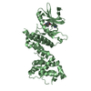
 Homo sapiens (human)
 X-RAY DIFFRACTION /
  Authors
 Citation
 Structure visualization
 Downloads & links
 Other downloads
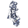


 PDBj
PDBj Assembly
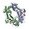
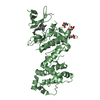

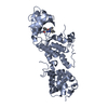
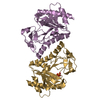
 Spodoptera frugiperda (fall armyworm)
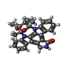
 Mass: 466.531 Da / Num. of mol.: 2 / Source method: isolated from a natural source / Formula: C28H26N4O3 / Comment: antibiotic*YM
 Mass: 18.015 Da / Num. of mol.: 58 / Source method: isolated from a natural source / Formula: H2O
 Sample preparation
 / Beamline: 21-ID-D / Wavelength: 0.97872 Å
 Processing