 Movie
Movie Controller


+ Open data

- Basic information
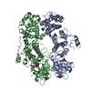
| Entry | Database: PDB / ID: 6w3a | ||||||
|---|---|---|---|---|---|---|---|
| Title | Structure of unphosphorylated IRE1 in complex with G-7658 | ||||||
 Components Components | Serine/threonine-protein kinase/endoribonuclease IRE1 | ||||||
 Keywords Keywords | TRANSFERASE / kinase / UPR | ||||||
| Function / homology |  Function and homology information Function and homology informationAIP1-IRE1 complex / Ire1 complex / mRNA splicing, via endonucleolytic cleavage and ligation / IRE1alpha activates chaperones / IRE1-TRAF2-ASK1 complex / insulin metabolic process / positive regulation of endoplasmic reticulum unfolded protein response / Hydrolases; Acting on ester bonds; Endoribonucleases producing 5'-phosphomonoesters / platelet-derived growth factor receptor binding / endothelial cell proliferation ...AIP1-IRE1 complex / Ire1 complex / mRNA splicing, via endonucleolytic cleavage and ligation / IRE1alpha activates chaperones / IRE1-TRAF2-ASK1 complex / insulin metabolic process / positive regulation of endoplasmic reticulum unfolded protein response / Hydrolases; Acting on ester bonds; Endoribonucleases producing 5'-phosphomonoesters / platelet-derived growth factor receptor binding / endothelial cell proliferation / nuclear inner membrane / IRE1-RACK1-PP2A complex / IRE1-mediated unfolded protein response / mRNA catabolic process / negative regulation of intrinsic apoptotic signaling pathway / intrinsic apoptotic signaling pathway in response to endoplasmic reticulum stress / cellular response to vascular endothelial growth factor stimulus / cellular response to unfolded protein / regulation of macroautophagy / positive regulation of vascular associated smooth muscle cell proliferation / endoplasmic reticulum unfolded protein response / RNA endonuclease activity / Hsp70 protein binding / response to endoplasmic reticulum stress / positive regulation of RNA splicing / cellular response to glucose stimulus / Hsp90 protein binding / ADP binding / positive regulation of JNK cascade / cellular response to hydrogen peroxide / : / protein phosphorylation / non-specific serine/threonine protein kinase / protein serine kinase activity / hydrolase activity / protein serine/threonine kinase activity / endoplasmic reticulum membrane / magnesium ion binding / enzyme binding / endoplasmic reticulum / protein homodimerization activity / mitochondrion / ATP binding / identical protein binding / cytoplasm Similarity search - Function | ||||||
| Biological species |  Homo sapiens (human) Homo sapiens (human) | ||||||
| Method |  X-RAY DIFFRACTION / SYNCHROTRON / MOLECULAR REPLACEMENT / Resolution: 2.606 Å X-RAY DIFFRACTION / SYNCHROTRON / MOLECULAR REPLACEMENT / Resolution: 2.606 Å | ||||||
 Authors Authors | Ferri, E. / Wang, W. / Joachim, R. / Mortara, K. | ||||||
 Citation Citation | Journal: Nat Commun / Year: 2020 Title: Activation of the IRE1 RNase through remodeling of the kinase front pocket by ATP-competitive ligands. Authors: Ferri, E. / Le Thomas, A. / Wallweber, H.A. / Day, E.S. / Walters, B.T. / Kaufman, S.E. / Braun, M.G. / Clark, K.R. / Beresini, M.H. / Mortara, K. / Chen, Y.A. / Canter, B. / Phung, W. / ...Authors: Ferri, E. / Le Thomas, A. / Wallweber, H.A. / Day, E.S. / Walters, B.T. / Kaufman, S.E. / Braun, M.G. / Clark, K.R. / Beresini, M.H. / Mortara, K. / Chen, Y.A. / Canter, B. / Phung, W. / Liu, P.S. / Lammens, A. / Ashkenazi, A. / Rudolph, J. / Wang, W. | ||||||
| History |
|
- Structure visualization
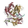
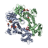
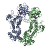
| Structure viewer | Molecule: MolmilJmol/JSmol |
|---|
- Downloads & links
-Download
| PDBx/mmCIF format | 6w3a.cif.gz | 411.8 KB | Display | PDBx/mmCIF format |
|---|---|---|---|---|
| PDB format | pdb6w3a.ent.gz | 339.4 KB | Display | PDB format |
| PDBx/mmJSON format | 6w3a.json.gz | Tree view | PDBx/mmJSON format | |
| Others |  Other downloads Other downloads |
-Validation report
| Arichive directory | https://data.pdbj.org/pub/pdb/validation_reports/w3/6w3aftp://data.pdbj.org/pub/pdb/validation_reports/w3/6w3a | HTTPS FTP |
|---|
-Related structure data
| Related structure data | 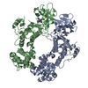 6w39C  6w3bC 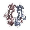 6w3cC  6w3eC  6w3kC 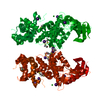 5hgiS S: Starting model for refinement C: citing same article ( |
|---|---|
| Similar structure data |


-Links
 PDBj
PDBj- Assembly
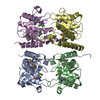
| Deposited unit | 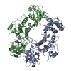
| ||||||||
|---|---|---|---|---|---|---|---|---|---|
| 1 |
| ||||||||
| Unit cell |
|
-Components
| #1: Protein | Mass: 49561.496 Da / Num. of mol.: 2 Source method: isolated from a genetically manipulated source Source: (gene. exp.) Homo sapiens (human) / Gene: ERN1, IRE1 / Cell line (production host): Sf9 / Production host:   Spodoptera frugiperda (fall armyworm) Spodoptera frugiperda (fall armyworm)References: UniProt: O75460, non-specific serine/threonine protein kinase, Hydrolases; Acting on ester bonds; Endoribonucleases producing 5'-phosphomonoesters #2: Chemical | 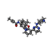  Mass: 492.572 Da / Num. of mol.: 2 / Source method: obtained synthetically / Formula: C29H28N6O2 / Feature type: SUBJECT OF INVESTIGATION Mass: 492.572 Da / Num. of mol.: 2 / Source method: obtained synthetically / Formula: C29H28N6O2 / Feature type: SUBJECT OF INVESTIGATION#3: Water | ChemComp-HOH / |  Mass: 18.015 Da / Num. of mol.: 42 / Source method: isolated from a natural source / Formula: H2O Mass: 18.015 Da / Num. of mol.: 42 / Source method: isolated from a natural source / Formula: H2OHas ligand of interest | Y | |
|---|
-Experimental details
-Experiment
| Experiment | Method: X-RAY DIFFRACTION / Number of used crystals: 1 |
|---|
- Sample preparation
| Crystal | Density Matthews: 2.63 Å3/Da / Density % sol: 53.2 % |
|---|---|
| Crystal grow | Temperature: 277 K / Method: vapor diffusion, sitting drop Details: 0.2 M magnesium formate, 20 %w/v PEG 3350, and 1% CYMAL-1 |
-Data collection
| Diffraction | Mean temperature: 100 K / Serial crystal experiment: N | ||||||||||||||||||||||||||||||
|---|---|---|---|---|---|---|---|---|---|---|---|---|---|---|---|---|---|---|---|---|---|---|---|---|---|---|---|---|---|---|---|
| Diffraction source | Source: SYNCHROTRON / Site: SSRL  / Beamline: BL12-2 / Wavelength: 0.97946 Å / Beamline: BL12-2 / Wavelength: 0.97946 Å | ||||||||||||||||||||||||||||||
| Detector | Type: DECTRIS PILATUS 6M / Detector: PIXEL / Date: Jan 31, 2018 | ||||||||||||||||||||||||||||||
| Radiation | Protocol: SINGLE WAVELENGTH / Monochromatic (M) / Laue (L): M / Scattering type: x-ray | ||||||||||||||||||||||||||||||
| Radiation wavelength | Wavelength: 0.97946 Å / Relative weight: 1 | ||||||||||||||||||||||||||||||
| Reflection | Resolution: 2.606→82.013 Å / Num. obs: 25334 / % possible obs: 91.5 % / Redundancy: 6.7 % / Biso Wilson estimate: 46.77 Å2 / CC1/2: 0.998 / Rmerge(I) obs: 0.148 / Rpim(I) all: 0.061 / Rrim(I) all: 0.16 / Net I/σ(I): 10.4 / Num. measured all: 170557 | ||||||||||||||||||||||||||||||
| Reflection shell | Diffraction-ID: 1
|
- Processing
| Software |
| ||||||||||||||||||||||||||||||||||||||||
|---|---|---|---|---|---|---|---|---|---|---|---|---|---|---|---|---|---|---|---|---|---|---|---|---|---|---|---|---|---|---|---|---|---|---|---|---|---|---|---|---|---|
| Refinement | Method to determine structure: MOLECULAR REPLACEMENT Starting model: 5HGI Resolution: 2.606→58.124 Å / SU ML: 0.37 / Cross valid method: THROUGHOUT / σ(F): 1.34 / Phase error: 35.38 / Stereochemistry target values: ML
| ||||||||||||||||||||||||||||||||||||||||
| Solvent computation | Shrinkage radii: 0.9 Å / VDW probe radii: 1.11 Å / Solvent model: FLAT BULK SOLVENT MODEL | ||||||||||||||||||||||||||||||||||||||||
| Displacement parameters | Biso max: 447.06 Å2 / Biso mean: 69.8148 Å2 / Biso min: 10.17 Å2 | ||||||||||||||||||||||||||||||||||||||||
| Refinement step | Cycle: final / Resolution: 2.606→58.124 Å
| ||||||||||||||||||||||||||||||||||||||||
| LS refinement shell | Resolution: 2.606→2.6991 Å
| ||||||||||||||||||||||||||||||||||||||||
| Refinement TLS params. | Method: refined / Origin x: 96.4536 Å / Origin y: -8.1553 Å / Origin z: 31.5024 Å
| ||||||||||||||||||||||||||||||||||||||||
| Refinement TLS group |
|
