 Movie
Movie Controller

[English] 日本語
 Yorodumi
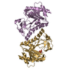
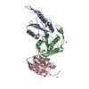
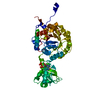
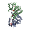
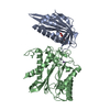
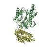
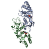
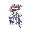
- PDB-6no1: ADP bound to K46bE mutant ATP-grasp fold of Blastocystis hominis ... -
+ Open data

- Basic information
| Entry | Database: PDB / ID: 6no1 | ||||||
|---|---|---|---|---|---|---|---|
| Title | ADP bound to K46bE mutant ATP-grasp fold of Blastocystis hominis succinyl-CoA synthetase | ||||||
 Components Components | Succinate--CoA ligase [ADP-forming] subunit beta | ||||||
 Keywords Keywords | LIGASE / Mutant / Complex | ||||||
| Function / homology |  Function and homology information Function and homology informationhydrogenosome / succinate-CoA ligase (ADP-forming) / succinate-CoA ligase complex / succinate-CoA ligase (ADP-forming) activity / succinyl-CoA metabolic process / tricarboxylic acid cycle / magnesium ion binding / mitochondrion / ATP binding Similarity search - Function | ||||||
| Biological species | Blastocystis sp. subtype 1 | ||||||
| Method |  X-RAY DIFFRACTION / SYNCHROTRON / Resolution: 2.44 Å X-RAY DIFFRACTION / SYNCHROTRON / Resolution: 2.44 Å | ||||||
 Authors Authors | Huang, J. / Fraser, M.E. | ||||||
| Funding support |  Canada, 1items Canada, 1items
| ||||||
 Citation Citation | Journal: Acta Crystallogr D Struct Biol / Year: 2019 Title: ATP-specificity of succinyl-CoA synthetase from Blastocystis hominis. Authors: Huang, J. / Nguyen, V.H. / Hamblin, K.A. / Maytum, R. / van der Giezen, M. / Fraser, M.E. | ||||||
| History |
|
- Structure visualization
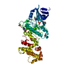
| Structure viewer | Molecule: MolmilJmol/JSmol |
|---|
- Downloads & links
-Download
| PDBx/mmCIF format | 6no1.cif.gz | 261.2 KB | Display | PDBx/mmCIF format |
|---|---|---|---|---|
| PDB format | pdb6no1.ent.gz | 216.3 KB | Display | PDB format |
| PDBx/mmJSON format | 6no1.json.gz | Tree view | PDBx/mmJSON format | |
| Others |  Other downloads Other downloads |
-Validation report
| Arichive directory | https://data.pdbj.org/pub/pdb/validation_reports/no/6no1ftp://data.pdbj.org/pub/pdb/validation_reports/no/6no1 | HTTPS FTP |
|---|
-Related structure data
| Related structure data | 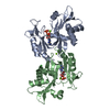 6no0C 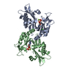 6no2C 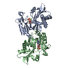 6no3C 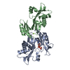 6no4C 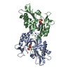 6no5C 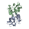 6no6C C: citing same article ( |
|---|---|
| Similar structure data |

-Links
 PDBj
PDBj


- Assembly
| Deposited unit | 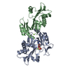
| ||||||||
|---|---|---|---|---|---|---|---|---|---|
| 1 |
| ||||||||
| Unit cell |
|
-Components
| #1: Protein | Mass: 27610.992 Da / Num. of mol.: 2 / Fragment: UNP residues 16-253 / Mutation: K46E Source method: isolated from a genetically manipulated source Source: (gene. exp.)  Blastocystis sp. subtype 1 (strain ATCC 50177 / NandII) (eukaryote) Blastocystis sp. subtype 1 (strain ATCC 50177 / NandII) (eukaryote)Strain: ATCC 50177 / NandII / Gene: SCSb / Production host:  References: UniProt: B3FHP0, succinate-CoA ligase (ADP-forming) #2: Chemical | ChemComp-ADP / |   Mass: 427.201 Da / Num. of mol.: 1 / Source method: obtained synthetically / Formula: C10H15N5O10P2 / Feature type: SUBJECT OF INVESTIGATION / Comment: ADP, energy-carrying molecule*YM Mass: 427.201 Da / Num. of mol.: 1 / Source method: obtained synthetically / Formula: C10H15N5O10P2 / Feature type: SUBJECT OF INVESTIGATION / Comment: ADP, energy-carrying molecule*YM#3: Chemical |   Mass: 24.305 Da / Num. of mol.: 2 / Source method: obtained synthetically / Formula: Mg Mass: 24.305 Da / Num. of mol.: 2 / Source method: obtained synthetically / Formula: Mg#4: Water | ChemComp-HOH / |  Mass: 18.015 Da / Num. of mol.: 20 / Source method: isolated from a natural source / Formula: H2O Mass: 18.015 Da / Num. of mol.: 20 / Source method: isolated from a natural source / Formula: H2OHas protein modification | Y | |
|---|
-Experimental details
-Experiment
| Experiment | Method: X-RAY DIFFRACTION / Number of used crystals: 1 |
|---|
- Sample preparation
| Crystal | Density Matthews: 2.37 Å3/Da / Density % sol: 48.03 % |
|---|---|
| Crystal grow | Temperature: 294 K / Method: vapor diffusion, hanging drop Details: 15% w/v PEG3350, 100 mM MES, pH 5.9, 125 mM ammonium tartrate, pH 8 |
-Data collection
| Diffraction | Mean temperature: 100 K / Serial crystal experiment: N | |||||||||||||||
|---|---|---|---|---|---|---|---|---|---|---|---|---|---|---|---|---|
| Diffraction source | Source: SYNCHROTRON / Site: CLSI / Beamline: 08ID-1 / Wavelength: 0.97949 Å | |||||||||||||||
| Detector | Type: DECTRIS PILATUS3 S 6M / Detector: PIXEL / Date: Feb 4, 2018 | |||||||||||||||
| Radiation | Monochromator: double crystal Si(111) / Protocol: SINGLE WAVELENGTH / Monochromatic (M) / Laue (L): M / Scattering type: x-ray | |||||||||||||||
| Radiation wavelength | Wavelength: 0.97949 Å / Relative weight: 1 | |||||||||||||||
| Reflection | Resolution: 2.44→80.12 Å / Num. obs: 20700 / % possible obs: 100 % / Redundancy: 6.2 % / Rmerge(I) obs: 0.107 / Net I/σ(I): 8 | |||||||||||||||
| Reflection shell |
|
- Processing
| Software |
| ||||||||||||||||||||||||||||||||||||||||||||||||||||||||||||||||||||||||||||||||||||||||||||||||||||||||||||||||||||||||||||||||||||||||||||||||||||||
|---|---|---|---|---|---|---|---|---|---|---|---|---|---|---|---|---|---|---|---|---|---|---|---|---|---|---|---|---|---|---|---|---|---|---|---|---|---|---|---|---|---|---|---|---|---|---|---|---|---|---|---|---|---|---|---|---|---|---|---|---|---|---|---|---|---|---|---|---|---|---|---|---|---|---|---|---|---|---|---|---|---|---|---|---|---|---|---|---|---|---|---|---|---|---|---|---|---|---|---|---|---|---|---|---|---|---|---|---|---|---|---|---|---|---|---|---|---|---|---|---|---|---|---|---|---|---|---|---|---|---|---|---|---|---|---|---|---|---|---|---|---|---|---|---|---|---|---|---|---|---|---|
| Refinement | Resolution: 2.44→69.235 Å / SU ML: 0.44 / Cross valid method: THROUGHOUT / σ(F): 1.44 / Phase error: 38.65
| ||||||||||||||||||||||||||||||||||||||||||||||||||||||||||||||||||||||||||||||||||||||||||||||||||||||||||||||||||||||||||||||||||||||||||||||||||||||
| Solvent computation | Shrinkage radii: 0.9 Å / VDW probe radii: 1.11 Å | ||||||||||||||||||||||||||||||||||||||||||||||||||||||||||||||||||||||||||||||||||||||||||||||||||||||||||||||||||||||||||||||||||||||||||||||||||||||
| Displacement parameters | Biso max: 273.11 Å2 / Biso mean: 101.9796 Å2 / Biso min: 31.8 Å2 | ||||||||||||||||||||||||||||||||||||||||||||||||||||||||||||||||||||||||||||||||||||||||||||||||||||||||||||||||||||||||||||||||||||||||||||||||||||||
| Refinement step | Cycle: final / Resolution: 2.44→69.235 Å
| ||||||||||||||||||||||||||||||||||||||||||||||||||||||||||||||||||||||||||||||||||||||||||||||||||||||||||||||||||||||||||||||||||||||||||||||||||||||
| LS refinement shell | Refine-ID: X-RAY DIFFRACTION / Rfactor Rfree error: 0 / Total num. of bins used: 7 / % reflection obs: 100 %
| ||||||||||||||||||||||||||||||||||||||||||||||||||||||||||||||||||||||||||||||||||||||||||||||||||||||||||||||||||||||||||||||||||||||||||||||||||||||
| Refinement TLS params. | Method: refined / Refine-ID: X-RAY DIFFRACTION
| ||||||||||||||||||||||||||||||||||||||||||||||||||||||||||||||||||||||||||||||||||||||||||||||||||||||||||||||||||||||||||||||||||||||||||||||||||||||
| Refinement TLS group |
|
