 Movie
Movie Controller

[English] 日本語
 Yorodumi
- PDB-6no3: ADP bound to V113bL mutant ATP-grasp fold of Blastocystis hominis... -
+ Open data

- Basic information
| Entry | Database: PDB / ID: 6no3 | ||||||
|---|---|---|---|---|---|---|---|
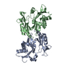
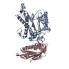
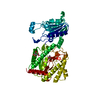
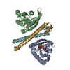
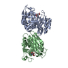
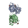
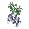
| Title | ADP bound to V113bL mutant ATP-grasp fold of Blastocystis hominis succinyl-CoA synthetase | ||||||
 Components Components | Succinate--CoA ligase [ADP-forming] subunit beta | ||||||
 Keywords Keywords | LIGASE / Complex / Mutant | ||||||
| Function / homology |  Function and homology information Function and homology informationhydrogenosome / succinate-CoA ligase (ADP-forming) / succinate-CoA ligase complex / succinate-CoA ligase (ADP-forming) activity / succinyl-CoA metabolic process / tricarboxylic acid cycle / magnesium ion binding / mitochondrion / ATP binding Similarity search - Function | ||||||
| Biological species | Blastocystis sp. subtype 1 | ||||||
| Method |  X-RAY DIFFRACTION / SYNCHROTRON / Resolution: 1.939 Å X-RAY DIFFRACTION / SYNCHROTRON / Resolution: 1.939 Å | ||||||
 Authors Authors | Huang, J. / Fraser, M.E. | ||||||
| Funding support |  Canada, 1items Canada, 1items
| ||||||
 Citation Citation | Journal: Acta Crystallogr D Struct Biol / Year: 2019 Title: ATP-specificity of succinyl-CoA synthetase from Blastocystis hominis. Authors: Huang, J. / Nguyen, V.H. / Hamblin, K.A. / Maytum, R. / van der Giezen, M. / Fraser, M.E. | ||||||
| History |
|
- Structure visualization
| Structure viewer | Molecule: MolmilJmol/JSmol |
|---|
- Downloads & links
-Download
| PDBx/mmCIF format | 6no3.cif.gz | 290.2 KB | Display | PDBx/mmCIF format |
|---|---|---|---|---|
| PDB format | pdb6no3.ent.gz | 236.8 KB | Display | PDB format |
| PDBx/mmJSON format | 6no3.json.gz | Tree view | PDBx/mmJSON format | |
| Others |  Other downloads Other downloads |
-Validation report
| Arichive directory | https://data.pdbj.org/pub/pdb/validation_reports/no/6no3ftp://data.pdbj.org/pub/pdb/validation_reports/no/6no3 | HTTPS FTP |
|---|
-Related structure data
| Related structure data |  6no0C  6no1C  6no2C  6no4C  6no5C  6no6C C: citing same article ( |
|---|---|
| Similar structure data |


-Links
 PDBj
PDBj


- Assembly
| Deposited unit | 
| ||||||||||||||||||||||||||||||||||||||||||||||||||||||||||||||||||||||||||||||||||||||||||
|---|---|---|---|---|---|---|---|---|---|---|---|---|---|---|---|---|---|---|---|---|---|---|---|---|---|---|---|---|---|---|---|---|---|---|---|---|---|---|---|---|---|---|---|---|---|---|---|---|---|---|---|---|---|---|---|---|---|---|---|---|---|---|---|---|---|---|---|---|---|---|---|---|---|---|---|---|---|---|---|---|---|---|---|---|---|---|---|---|---|---|---|
| 1 |
| ||||||||||||||||||||||||||||||||||||||||||||||||||||||||||||||||||||||||||||||||||||||||||
| Unit cell |
| ||||||||||||||||||||||||||||||||||||||||||||||||||||||||||||||||||||||||||||||||||||||||||
| Noncrystallographic symmetry (NCS) | NCS domain:
NCS domain segments: Ens-ID: 1
|