 Movie
Movie Controller

[English] 日本語
 Yorodumi
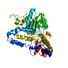
- PDB-4rtm: Complex of Escherichia coli DNA Adenine Methyltransferase (DAM) w... -
+ Open data

- Basic information
| Entry | Database: PDB / ID: 4rtm | ||||||
|---|---|---|---|---|---|---|---|
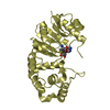
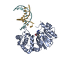
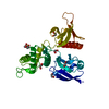
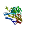
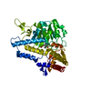
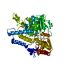
| Title | Complex of Escherichia coli DNA Adenine Methyltransferase (DAM) with AdoMet and with DNA Containing Distal Pap Regulon Sequence | ||||||
 Components Components |
| ||||||
 Keywords Keywords | Transferase/DNA / DAM METHYLATION / GATC RECOGNITION / BASE FLIPPING / BACTERIAL VIRULENCE / methylation-independent transcriptional repressor / Transferase-DNA complex | ||||||
| Function / homology |  Function and homology information Function and homology informationbacterial-type DNA replication initiation / site-specific DNA-methyltransferase (adenine-specific) / site-specific DNA-methyltransferase (adenine-specific) activity / S-adenosyl-L-methionine binding / DNA restriction-modification system / mismatch repair / response to UV / DNA-templated DNA replication / methylation / sequence-specific DNA binding Similarity search - Function | ||||||
| Biological species |  synthetic construct (others) | ||||||
| Method |  X-RAY DIFFRACTION / SYNCHROTRON / MOLECULAR REPLACEMENT / Resolution: 2.5 Å X-RAY DIFFRACTION / SYNCHROTRON / MOLECULAR REPLACEMENT / Resolution: 2.5 Å | ||||||
 Authors Authors | Horton, J.R. / Cheng, X. | ||||||
 Citation Citation | Journal: Nucleic Acids Res. / Year: 2015 Title: Structures of Escherichia coli DNA adenine methyltransferase (Dam) in complex with a non-GATC sequence: potential implications for methylation-independent transcriptional repression. Authors: Horton, J.R. / Zhang, X. / Blumenthal, R.M. / Cheng, X. | ||||||
| History |
|
- Structure visualization
| Structure viewer | Molecule: MolmilJmol/JSmol |
|---|
- Downloads & links
-Download
| PDBx/mmCIF format | 4rtm.cif.gz | 81.8 KB | Display | PDBx/mmCIF format |
|---|---|---|---|---|
| PDB format | pdb4rtm.ent.gz | 56.4 KB | Display | PDB format |
| PDBx/mmJSON format | 4rtm.json.gz | Tree view | PDBx/mmJSON format | |
| Others |  Other downloads Other downloads |
-Validation report
| Arichive directory | https://data.pdbj.org/pub/pdb/validation_reports/rt/4rtmftp://data.pdbj.org/pub/pdb/validation_reports/rt/4rtm | HTTPS FTP |
|---|
-Related structure data
| Related structure data | 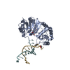 4rtjC 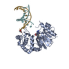 4rtkC 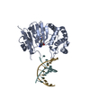 4rtlC  4rtnC 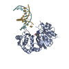 4rtoC  4rtpC 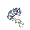 4rtqC 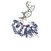 4rtrC 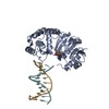 4rtsC 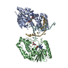 2g1pS S: Starting model for refinement C: citing same article ( |
|---|---|
| Similar structure data |


-Links
 PDBj
PDBj


- Assembly
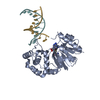
| Deposited unit | 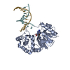
| ||||||||
|---|---|---|---|---|---|---|---|---|---|
| 1 |
| ||||||||
| Unit cell |
|
-Components
| #1: Protein | Mass: 34330.961 Da / Num. of mol.: 1 Source method: isolated from a genetically manipulated source Source: (gene. exp.) |
|---|---|
| #2: DNA chain | Mass: 3357.223 Da / Num. of mol.: 1 / Source method: obtained synthetically / Source: (synth.) synthetic construct (others) |
| #3: DNA chain | Mass: 3348.209 Da / Num. of mol.: 1 / Source method: obtained synthetically / Source: (synth.) synthetic construct (others) |
| #4: Chemical | ChemComp-SAM /   Mass: 398.437 Da / Num. of mol.: 1 / Source method: obtained synthetically / Formula: C15H22N6O5S Mass: 398.437 Da / Num. of mol.: 1 / Source method: obtained synthetically / Formula: C15H22N6O5S |
| #5: Water | ChemComp-HOH /  Mass: 18.015 Da / Num. of mol.: 50 / Source method: isolated from a natural source / Formula: H2O Mass: 18.015 Da / Num. of mol.: 50 / Source method: isolated from a natural source / Formula: H2O |
| Sequence details | AUTHOR STATES THAT RESIDUES 175 WERE MODELED AS SER INSTEAD OF ALA BECAUSE OF EXTRA DENSITY. |
-Experimental details
-Experiment
| Experiment | Method: X-RAY DIFFRACTION / Number of used crystals: 1 |
|---|
- Sample preparation
| Crystal | Density Matthews: 2 Å3/Da / Density % sol: 38.55 % |
|---|---|
| Crystal grow | Temperature: 289 K / Method: vapor diffusion, hanging drop / pH: 6.8 Details: 21% PEG200, 100mM KCl, 10mM MgSO4, 100mM MES buffer, pH 6.8, VAPOR DIFFUSION, HANGING DROP, temperature 289K |
-Data collection
| Diffraction | Mean temperature: 100 K |
|---|---|
| Diffraction source | Source: SYNCHROTRON / Site: APS  / Beamline: 22-ID / Wavelength: 1 Å / Beamline: 22-ID / Wavelength: 1 Å |
| Detector | Type: MARMOSAIC 300 mm CCD / Detector: CCD / Date: Dec 15, 2006 |
| Radiation | Monochromator: Si(111) / Protocol: SINGLE WAVELENGTH / Monochromatic (M) / Laue (L): M / Scattering type: x-ray |
| Radiation wavelength | Wavelength: 1 Å / Relative weight: 1 |
| Reflection | Resolution: 2.5→32.431 Å / Num. all: 12050 / Num. obs: 12050 / % possible obs: 100 % / Observed criterion σ(F): -3 / Observed criterion σ(I): -3 / Redundancy: 12 % / Biso Wilson estimate: 41.8 Å2 / Rmerge(I) obs: 0.113 / Net I/σ(I): 10.7 |
| Reflection shell | Resolution: 2.5→2.58 Å / Redundancy: 12.9 % / Rmerge(I) obs: 0.439 / Mean I/σ(I) obs: 8.3 / Num. unique all: 1194 / % possible all: 100 |
- Processing
| Software |
| |||||||||||||||||||||||||||||||||||
|---|---|---|---|---|---|---|---|---|---|---|---|---|---|---|---|---|---|---|---|---|---|---|---|---|---|---|---|---|---|---|---|---|---|---|---|---|
| Refinement | Method to determine structure: MOLECULAR REPLACEMENT Starting model: PDB entry 2G1P Resolution: 2.5→32.431 Å / SU ML: 0.29 / σ(F): 1.35 / Phase error: 23.33 / Stereochemistry target values: ML
| |||||||||||||||||||||||||||||||||||
| Solvent computation | Shrinkage radii: 0.9 Å / VDW probe radii: 1.11 Å / Solvent model: FLAT BULK SOLVENT MODEL | |||||||||||||||||||||||||||||||||||
| Refinement step | Cycle: LAST / Resolution: 2.5→32.431 Å
| |||||||||||||||||||||||||||||||||||
| Refine LS restraints |
| |||||||||||||||||||||||||||||||||||
| LS refinement shell |
|
